

Review Article
Volume-1 Issue-1, 2021
The effect of Wheatgrass and Zinc on P53, APC, Bcl2, Bax expression in Rat colon cancer
Received Date: October 15, 2022
Accepted Date: November 15, 2022
Published Date: November 17, 2022
Journal Information
Abstract
Background: Colorectal cancer (CRC) is the most frequent type of gastrointestinal cancer. The present study explores role of wheatgrass and zinc as preventive therapy.
Setting and Design: Forty-eight male rats divided into eight groups were given, diet with 1,2 Dimethylhydrazine (DMH), Zinc, wheatgrass, wheatgrass + zinc, DMH with zinc, DMH with wheatgrass, and DMH with a combination of zinc and wheatgrass. One group served as control. Protein expressions of p53, B-cell lymphoma-2 (Bcl-2), BCL2 Associated X (Bax) and adenomatous polyposis coli (APC) proteins were investigated using immunohistochemistry.
Results: Administration of DMH led to increased protein expression of p53 and Bcl-2 and reduced expression of APC and Bax. Zinc and wheatgrass supplementation was found to decrease Bcl-2 and p53 expression and increase expression of Bax and APC proteins.
Conclusion: Study demonstrated that zinc and wheatgrass in combination are effective in bringing about a delay in the chain of molecular events leading to onset and progression of DMH-induced colon carcinogenesis in experimental rats. Results point towards possible use of chemo preventive regimens as an adjunct treatment in CRC.
Key words
Wheatgrass; Zinc; P53; APC; Bcl-2; Bax;
Abbreviations: “CRC (Colorectal cancer)”, “Zn (Zinc)”, “DMH (1,2 Dimethyl hydrazine)”, “APC (Adenomatous Polyposis Coli)”, “Bcl-2 (B-cell lymphoma-2)”, “EDTA (Ethylenediaminetetracetic acid)”, “FaSL (Fas ligand)”, “IHC(Immunohistochemistry)”, “K-ras (Kirsten-ras)”, “μMMicromolar”, “SD (Sprague Dawley)”.
Groups |
Intervention |
Dose schedule |
Group 1 (Control) |
Standard laboratory feed, water ad libitum |
During the period of experimentation |
Group2 (DMH) |
DMH in 1mM EDTA-saline dose of 30mg/kg body weight. (19) |
Weekly injection of DMH for 16 weeks. |
Group3 (DMH+Zn) |
DMH in 1mM EDTA- saline dose of 30mg/ kg body weight + 227mg/l of Zn (zinc sulfate |
Weekly injection of DMH for 16 weeks. Zn dose was started 15 days before the first injection of DMH. |
Group4 (DMH+WG) |
DMH in 1mM EDTA-saline dose of 30mg/ kg body wt. + 100mg/kg body wt. of WG in drinking water. |
Weekly injection of DMH for 16 weeks and WG given orally daily and was started 15 days before DMH injection. |
Group5 (DMH+WG+Zn) |
DMH in 1mM EDTA-saline dose of 30mg/kg body wt. + Zn (227mg/l) + WG (100mg/kg body wt). |
Weekly injection of DMH for 16 weeks. WG and Zn dose was started 15 days before the first injection of DMH. |
Group6 (Zn alone) |
Zn (227mg/l) in drinking water. |
Daily Zn alone for 16 weeks. |
Group7 (WG alone) |
Tablets of WG (100mg/kg body wt.) in drinking water. |
Daily WG alone was given orally, for 16 weeks |
Group 8 (Zn+ WG) |
Tablets of WG (100mg/kg body wt.) daily. + Zn (227mg/l) in drinking water |
Daily WG and Zn were given for 16 weeks. This dose was started 15 days before the first injection of DMH |
Score |
0 |
1 |
2 |
3 |
4 |
Nuclear Postive Cells |
<10% |
10-25% |
25-50% |
50-75% |
>75% |
Cytoplasmic Positive Cells |
Negative |
Few Nuclei |
10% |
10-50 % |
>50% |
Intensity Score |
1 |
2 |
3 |
Intensity Staining |
Weak Staining |
Moderate Staining |
Strong Staining |
|
Control Group 1 |
DMH |
DMH+Zn |
DMH+WG |
DMH+WG+Zn |
Zn Group 6 |
WG |
Zn+ WG |
Nuclear Positivity score |
0 |
3 |
2 |
2 |
1 |
0 |
0 |
0 |
Nuclear |
2 |
1 |
1 |
1 |
2 |
2 |
2 |
2 |
Cytoplasmic positivity score |
0 |
2 |
2 |
1 |
0 |
0 |
0 |
0 |
Cytoplasmic |
2 |
0 |
1 |
1 |
2 |
2 |
2 |
2 |
Intensity Score (p53), APC, |
0,1,0,2 |
1,1,1,1 |
1,2,1,1 |
1,2,1,1 |
1,1,1,2 |
0,1,1,2 |
0,1,1,2 |
0,1,1,2 |
IHC Score (P53), APC, |
0,2,0,4 |
9,1,2,0 |
2,2,1,1 |
2,2,1,1 |
1,2,0,4 |
0,2,0,4 |
0,2,0,4 |
0,2,0,4 |
DMH: Dimethylhydrazine, WG: Wheatgrass, Zn: Zinc, IHC(Immunohistochemistry)
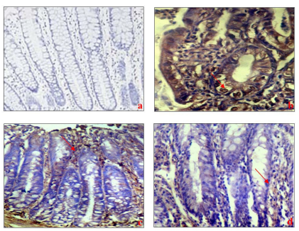 |
| Figure 1: Represents the immunohistochemical staining of p53 in colon tissue of a) Control animals showing negative nuclear staining. b) DMH group showing highly expressed p53 nuclear positivity. c) DMH+Zn group showing weak nuclear positivity. d) DMH+WG group showing weak nuclear positivity. e) DMH+Zn+WG group showing negative nuclear staining. f) Zn+WG group showing negative nuclear staining (X 20). DMH: Dimethylhydrazine, WG: Wheatgrass, Zn: Zinc |
 |
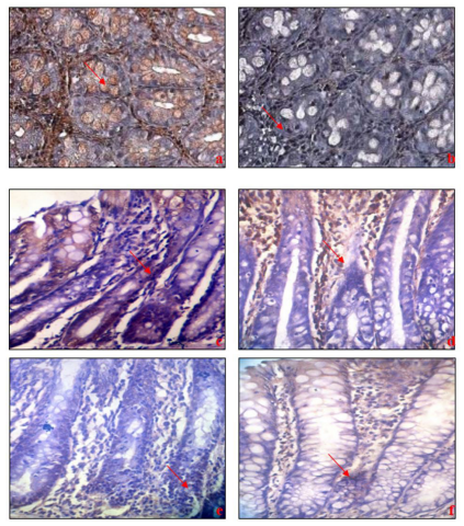 |
| Figure 3:Represents the immunohistochemical staining of APC in colon tissue of a) Control animals showing moderate nuclear staining. b) DMH group showing negative nuclear staining. c) DMH+Zn group showing weak nuclear positivity, expression increased as compared to DMH. d) DMH+WG group showing weak nuclear positivity, expression was more nuclear positive than DMH+Zn. e) DMH+Zn+WG group showing moderate nuclear positivity. f) Zn+WG group showing staining similar to control (X 20). DMH: Dimethylhydrazine, WG: Wheatgrass, Zn: Zinc |
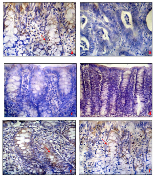 |
| Figure 4: Represents the immunohistochemical staining of Bax in colon tissue of a) control animals showing moderate cytoplasmic positivity. b) DMH group showing negative cytoplasmic staining. c) DMH+Zn group showing weak cytoplasmic positivity. d) DMH+WG group showing weak cytoplasmic positivity. e) DMH+Zn+WG group showing moderate cytoplasmic positivity, comparable to control. f) Zn+WGM group showing moderate cytoplasmic positivity (X 20). DMH: Dimethylhydrazine, Zn: Zinc, WG: Wheatgrass |
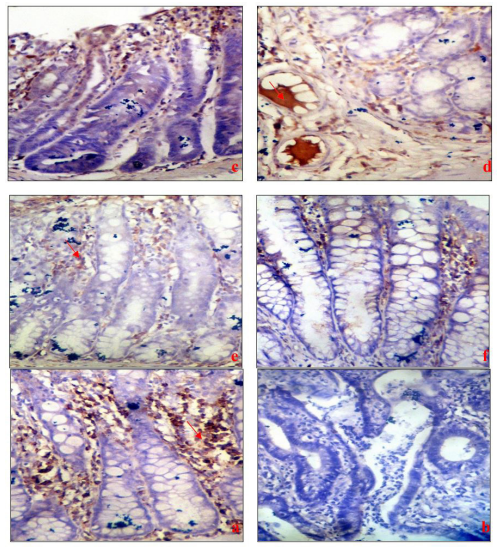 |
| Figure 5: Represents the immunohistochemical staining of Bcl2 in colon tissue of a) control animals showing negative cytoplasmic staining. b) DMH group showing weak cytoplasmic positive staining. Endothelial &lymphoid stained positive with no endothelial positivity. c) DMH+Zn group showing weak cytoplasmic positivity. d) DMH+WG group showing weak cytoplasmic positivity. e) DMH+Zn+WG group showing moderate cytoplasmic positivity, comparable to control. f) Zn+WG group showing negative cytoplasmic staining (X 20). DMH: Dimethylhydrazine, Zn: Zinc, WG: Wheatgrass |
Introduction
Unchecked division of abnormal cells characterizes cancer, when it occurs in the colon or rectum, it is called CRC [1]. In Western Countries quarter of deaths due to cancer [2]. Colon cancer ranks second as a cause of cancer deaths in the United States itself when men and women are combined [3]. People with low socioeconomic status (SES) have higher rates of incidence for CRC [4]. Within Asia, wide variations are seen in the incidence rates of CRC. It was found to be uniformly low in all south Asian countries and much higher in all developed Asian countries [5]. Colon cancer prevention integrates lifestyle factors and multistep genetic alterations [6]. The sporadic Colorectal tumors originate mostly from polyps or premalignant precursor lesions eventually progressing to clinically relevant tumors [6]. The sequence of events for this progression rests on mutation on APC protein and TP53 genes [6]. Apart from these, it is involving DNA repair or mismatch repair genes (MSH2, MLH1, PMS1, PMS2, and MSH6), the proto-oncogene K-ras (Kirsten-ras), DCC protein (deleted in CRC) [7]. BAX/BAK activation leads to apoptosis, and high levels of BCL-XL are found in CRC lesions, thus making it a prime interest target for cancer treatment [7-8].
Wheatgrass, barley grass, alfa-alfa cereal grasses have documented effects on boosting health and vitality [9]. Wheatgrass extract is also known to inhibit carcinogenesis [11]. This cereal grass is rich in chlorophyll, vitamins and enzymes, contains more than 60% chlorophyll, an active factor [10]. Some proponents claim that the Wheatgrass has an inhibiting effect on the proliferation of oral cancer cell lines and also it strengthens the immune system [12-13]. Zinc can induce apoptosis of human melanoma cells while increasing intracellular ROS and modulating p53 and FASL protein expression and also showed that zinc concentrations ranging from 33.7 μM to 75 μM Zn2+ induced apoptosis in the human melanoma cell line WM 266-4 [14]. Bax/Bcl-2 ratio expression is a prognostic marker in colorectal tumors compared to expression levels of Bax and/or Bcl-2 genes alone [15]. Expression pattern of p53, Bcl-2, and Annexin V in human CRC pre and post neo adjuvant FOLFOX chemotherapy showed that these contribute to apoptotic activities [16]. It is implicated in the activation and structural stabilization of p53, and in the caspase family of proteases activation, both having role in apoptosis [17]. Zinc deficiency promotes tumorigenesis in both the small intestine and colon [18]. Although zinc is a widely explored micronutrient, the dietary benefits of wheatgrass remain unexplored. No work has yet been done to study the antioxidant properties of wheatgrass and the combined effect of zinc & wheatgrass on the prevention of colon cancer. The role of wheatgrass as an alternative therapy in colon cancer needs to be explored in an experimental model before it is recommended for clinical use.
Moreover, there is the possibility of exploring the probable role of wheatgrass and zinc prophylactic therapy in delaying the initiation of events of colon tumor development, by controlling expressions of proteins involved in the apoptotic pathway. This study aimed at providing this initial evidence.
Animals
Male SD (Sprague Dawley) rats weighing 220-270g were used in this investigation, and they were acclimatised for one week before the start of experiments. Kept in polypropylene cages under standard conditions, rats were maintained as per the guidelines of the ethical committee of animal care of the Institute and following Indian laws.
Experimental design:
p53 Pathway and Apoptotic Studies
The role of wheatgrass and zinc in protein expressions of p53 and apoptotic pathway was studied by using immunohistochemistry (IHC). The antibody is used in low dilution in IHC & one needs only paraffin fixed tissue of the target area. It is advantageous over western blotting (WB) as higher dilution used in WB may sometimes result in non-detection of protein of interest.
Scoring of Immunostaining:
A scoring system was followed for Immunohistochemistry, based on previous study(21). The percentage positivity of tumour cells showing positive immunostaining and the intensity score as follows:
IHC Score (H) = % age positivity (P) X Intensity Score (S) The IHC Score ranged from 0-12 units
Scoring of Immunohistochemistry was done by the scoring system. The percentage positivity of tumor cells showing positive immunostaining was recorded.
Statistical Analysis
scoring system All data were tabulated as a number, percentage, and Immunohistochemistry was done using a.
Results
Immunohistochemistry of P53-in case of control and treated groups following 16 weeks of DMH treatment
Sections of normal and tumor areas were stained with a polyclonal antibody directed against p53. p53 immunoreactivity was not observed in normal colonic crypt cells of rats from any of the four groups (group 1, 6, 7 & 8) and IHC score=0 (Table 3).
Eight experimental groups each consisting of six male SD rats were given standard diets and divided in following 8 groups.
Although there was considerable variability in staining seen with the individual antibodies (Abcam laboratories, Thermo scientific, Genex bio) (Figure 1a-f). The staining was confined to the nuclei of the neoplastic cells and no cytoplasmic staining was detected by any of the monoclonal antibodies (Dako laboratories) used. The staining within the tumour nuclei was homogeneous and evenly distributed, although sometimes with an accentuation around the nuclear membrane as seen in the DMH group in which p53 was highly expressed (IHC score= 9). Very weak nuclear expression was seen in zinc and wheatgrass supplemented groups along with DMH treatment. However, nuclear negativity was observed with a combined supplementation of zinc and wheatgrass to DMH-treated rats as apoptosis increased in these groups. It was scored as positive when at least 10% of the tumour nuclei showed positivity. Using this criterion, 4 out of 6 animals were found to be immunohistochemically positive for p53 protein in the DMH group and two animals showed weak nuclear positivity. Zinc and wheatgrass individual supplementation along with DMH showed weak nuclear positivity as very few cells were found positive (IHC sore=2). Synergistic ingestion of zinc and wheatgrass showed negative nuclear staining (IHC score=1).
Immunohistochemistry of APC-in case of control and treated groups following 16 weeks of DMH treatment
APC protein was scored as positive when at least 10% of the tumour nuclei showed positivity. Using this criterion, 1 out of 6 animals were found to be immunohistochemically positive for APC protein in the DMH group and 5 animals showed weak nuclear positivity. As shown in (Fig 2) moderate nuclear staining was found in the control group (IHC score=2) as compared to the DMH group where APC was found weakly expressed as less than 10 nuclei were found positive (IHC score=1). However, levels of APC expression were regulated with zinc and wheatgrass supplementation as their expression became better in these groups and IHC scores increased to 2 in each case (Fig 2a-f). Zinc and wheatgrass when ingested alone showed weak expression of APC as compared to the control group. But when they were given in combination, synergistic action showed better results, and nuclear staining was almost found to be better than in the DMH group (IHC score=2) which confirmed the increased expression of APC in group5(Table 3).
Immunohistochemistry of Bax- in case of control and treated groups following 16 weeks of DMH treatment
Apoptosis-related genes ate subcategorized into proapoptosis genes and anti-apoptosis genes. Bcl-2 represses apoptosis, while Bax promotes apoptosis.
According to results, expression was scored as positive when at least 10% of the cells showed cytoplasmic positivity (Table 3). Using this criterion, all animals were found to be immunohistochemically positive for Bax protein in control. Only 2 animals showed weak cytoplasmic positivity in the DMH group. Control was found moderately positive as Bax showed cytoplasmic positivity in control as IHC score was 4 (Fig 3a-f). Expression was negative in the DMH group as no cytoplasmic staining was observed which confirmed the negative expression of Bax protein in this group). Weak positivity was observed in zinc and wheatgrass supplemented group (IHC score was 1 as weak cytoplasmic staining was observed). When zinc and wheatgrass were given in combination, strong cytoplasmic positivity was seen as IHC score increased to 4 as compared to the DMH group indicating the increased level of Bax protein. Thus, Cytoplasmic positivity was found similar to control rats in zinc and wheatgrass treated groups. These results indicated that the expression of Bax may have a role in intestinal adenocarcinoma.
Immunohistochemistry of Bcl2- in case of control and treated groups following 16 weeks of DMH treatment
The nuclear envelope or cell membrane and endoplasm contained Bcl-2 protein. Although the base of crypts in the normal small intestine expressed more Bcl-2, it was sometimes also present in the epithelial cells present on the surface. Bcl-2 could be detected extensively carcinoma cells nuclear envelope, and cell membrane but in comparison with protein Bax the intensity was relatively lower.
Expression was scored as positive when at least 10% of the cells showed cytoplasmic positivity (table 3). Using this criterion, only 1 animal was found to be immunohistochemically positive for Bcl2 protein in control. 4 out of 6 animals showed very weak cytoplasmic positivity in the DMH group (Figure 4a-f).
Bcl2 showed cytoplasmic negativity in the control group whereas positive expression was higher in colon changes induced by DMH. The cytoplasmic positivity was weak in zinc and wheatgrass supplemented groups (IHC score= 1) as Bcl2 expression was weak. It was also observed that expression was downregulated and only 1 animal showed weak cytoplasmic positivity on combined ingestion of zinc and wheatgrass. No change was found when normal rats were supplemented with zinc and wheatgrass without any carcinogen.
Discussion
CRC is one of the leading causes of cancer death in both men and women [2]. The role of zinc is extensively studied as it has high antioxidant potential [22]. Although zinc is a wellstudied common nutrient, the dietary benefits of wheatgrass remain unexplored which is a medicinally important plant. Several different classes of compounds of pharmacological importance such as flavonoids and chlorophyll are present in wheatgrass extracts [23]. The present study aimed at evaluating the potential synergistic activity of zinc and wheatgrass utilizing immunohistochemical expression of tumour suppressor p53 and apoptotic proteins like APC, BCl2, and Bax. Wheatgrass has a place in the traditional medicine system for the treatment of various ailments, with its anticancer activity reported in vitro using cell lines like Hep2, Hela, HCT-15, and K562 without cytotoxic potential [24-25]. It was shown that wheatgrass extracts have Cytotoxicity and apoptosis activity, which inhibits proliferation of leukemia cells, and induces apoptosis. It was found a novel therapeutic approach for the treatment of Cancer [26]. One mechanism by which zinc has protective effects is ascribed to its collagen accumulation reducing property. The possible mechanism could be that post-translational modulation of p53 by zinc and wheatgrass supplementation has resulted in induction of apoptosis, which caused moderation in cell proliferation [27]. In the present study, DMH administration to rats for 16 weeks resulted in 100% of tumour incidence and tumour multiplicity in the colon of rats which is significantly high as compared to control rats. On a molecular basis, a modified inactive form of tumour suppressor p53 by DMH as revealed in the expression studies could have resulted in increased cell proliferation by causing a decline in apoptosis [28].
Immunohistochemical studies
the most important mechanism utilized by the body to remove cancerous cells is the triggering of apoptosis via caspase cascade. Alterations in the apoptosis process on the addition of zinc and wheatgrass to diets of DMH-treated animals were found in the current study.
p53
can be found in the serum of CRC [31]. Therefore, detection of protein usually by the immune-histochemical technique can be currently used as an indirect indicator of mutations in p53. Overexpressed p53 protein is very common in human malignancies. In the current study, post DMH treatment protein expression of p53 was increased. Occasional inflammatory cells infiltrating the tumour were also positive. The increase in p53 expression observed is a consequence of the triggering of natural defenses of the cell against external stimuli/carcinogens [32]. However, p53 activity is kept low in normal cells as the cell cycle is not disrupted and cells do not undergo untimely death. In the case of DNA damage, the p53-mediated pathways are activated leading to cell cycle arrest and repair of the DNA.P53 a tumour suppressor is highly regulated acting as a cell-cycle blocker that promotes apoptosis. Its expression is induced by various stress stimuli including oncogene activation and DNA damage [29]. The TP53 gene commonly mutated in CRC is significantly involved in the control of the cell cycle and apoptosis [30]. Mutations in p53 are proposed to be relatively late events in the development of colorectal tumours. Mutated TP53 is more commonly detected early in the sera and other body fluids. DNA containing mutations in TP53, the observed increase in the expression of p53 (number of nuclear positive cells) is the result of the accumulation of modified inactive form of p53 which was unable to suppress tumour development as evidenced by an increased tumour incidence and tumour multiplicity in the DMH treated group. In this present study, the supplementation of zinc and wheatgrass separately or concomitantly brought about a significant decrease in the p53expression in tissue samples as nuclear positivity decreased in these groups. The observed decrease in the number of nuclear positive cells in immunostaining of p53 might be the result of the synergistic modulatory effect of zinc and wheatgrass on p53 to improve its tumour suppressor activity, which was confirmed by a decline in the tumour incidence and tumor multiplicity in these groups. P53 induced cell cycle arrest, via its influence downstream effectors of transcription, gives the ability to cells to repair genomic damage before critical stages of DNA synthesis and mitosis [33]. The functional conformation of the p53 protein is affected by zinc and the addition of zinc mediates the reversion to wild-type p53 [34]. On further addition of metal-chelating reagents reverts conformational change from wild type to the mutant of p53.
APC
In the present study, about 4 out of 6 animals showed a reduction in APC expression in the tissue of DMH treated rats was found suggesting that APC expression decreases during the shift from normal epithelium to carcinoma. Similar to our findings, [35] reported reduced APC expression in 34.7 % of the tumor tissues. [36] However, reported 83 % of colon cancer had a loss of APC expression.
Apoptotic proteins: Bcl-2 and Bax
Certain genes control apoptosis acting as death switches. The most essential regulator is the Bcl-2 gene family reported in neoplasms of breast, prostate, and thyroid carcinoma, and smallcell and large-cell carcinoma [37]. Among apoptosis-related proteins, Bax was the first protein isolated and it had homology (throughout two highly conserved regions) with Bcl-2 [38]. In the present study, the Bcl-2 protein expression showed a significant increase while that of Bax showed a significant expression decrease in the DMH treated rat colons. Bcl2 is an anti-apoptotic protein [29], its expression increase indicates declined apoptosis. Bax acts as a critical checkpoint in the process of apoptosis in epithelial cells and is crucial regarding oncogenesis and cancer therapy [39]. It has been reported that the majority of tumours were Bax- expression dominated and with weak positivity of both Bcl-2 and Bcl-xL. This is in contrast to a study in which overexpression of all the three proteins has been reported [40]. Zinc and wheatgrass treatments individually, as well as given, concomitantly were successful in inducing apoptosis in DMH treated rats by controlling the expression of Bcl2 and Bax protein. On treatment with zinc and wheatgrass, there was an escalation of Bax expression and reduction in Bcl-2 expression as compared to the DMH group. The primary reason is the stabilization of the active tumour suppressor form of p53. Secondly, these nutrients have also been reported to contribute towards the maintenance of genome stability [17] and chromatin remodeling encompassing events that play an important role in cancer progression [41]. Fermented wheat aleurone layer was detected beneficial subfraction, with fermentation metabolites inducing apoptosis in human colon adenocarcinoma cell lines [42].
In the present study, it is demonstrated that supplementing zinc and wheatgrass synergistically can induce apoptosis by preserving the Bcl2-to-Bax ratio in rapidly proliferating cells. Considering the growing evidence for the chemopreventive action of wheatgrass and dietary chlorophyll, the lack of evidence uncovering insights into chlorophyll as a chemoprotective agent is surprising. It is imperative to continue molecular and biochemical level investigations compiling a database of foods high in phytochemicals. Wheatgrass also has similar antioxidant potential as zinc in maintaining the ultra-structural balances of the rat colon subjected to DMH treatment.
Therefore, the primary goal of this study was to determine whether the synergistic ingestion of zinc and wheatgrass could prevent tumour formation. As 70% of microvilli was found to be intact in this group with minimal cellular changes. The intact mitochondria structure also provides cytological evidence of efficient p53 as inactive p53 leads to premature mitochondrial senescence [43]. This can provide evidence that zinc and wheatgrass may have chemopreventive potential against CRC by maintaining cellular integrity
Acknowledgments
Thanks to Central library AIIMS Rishikesh for the plagiarism check.
Funding support
This work was supported by Indian Council of Medical Research vide no 3/2/2/49/2010/NCD-III. and Author prof. Satyavati Rana has received research from PGIMER Chandigarh.
References
- American Cancer Society (2020) Colorectal Cancer Facts & Figures 2020-2022. Atlanta: American Cancer Society
- Landis SH, Murray T, Bolden S, Wingo PA (1999) Cancer Statistics. Cancer J Clin 49: 8-31.
- Islami F, Sauer AG, Miller KD, Siegel RL, Fedewa SA et al. (2018) Proportion and number of cancer cases and deaths attributable to potentially modifiable risk factors in the United States. CA Cancer J Clin 68: 31-54.
- Carethers JM, Doubeni CA (2020) Causes of Socioeconomic Disparities in Colorectal Cancer and Intervention Framework and Strategies. Gastroenterology 158: 354-67.
- Cunningham D, Atkin W, Lenz HJ, Lynch HT, Minsky B et al (2010) Colorectal cancer. Lancet 375: 1030-47.
- Ydy LRA, Camarço -Silva WR, Medeiros-Filho WV (2019) A Genetic Perspective on Colorectal Cancer Progression. In: Jeong, K., editor. Multidisciplinary Approach for Colorectal Cancer.
- Ramesh P, Medema JP (2020) BCL-2 family deregulation in colorectal cancer: potential for BH3 mimetics in therapy. Apoptosis 25: 305-20.
- Malhotra P, Anwar M, Nanda N, Kochhar R, Wig JD et al (2013) Alterations in K-ras, APC and p53-multiple genetic pathway in colorectal cancer among Indians. Tumour Biol 34: 1901-11.
- Gonzalez CA, Pera G, Agudo (2006) A Fruit and vegetables intake and the risk of stomach and oesophagus adenocarcinoma in the European Prospective Investigation into Cancer and Nutrition. International Journal of Cancer 118: 2559-66.
- Manju V, Nalini N (2005) Chemopreventive potential of luteolin during colon carcinogenesis induced by 1,2-dimethylhydrazine. Ital J Biochem 54: 268-75.
- Rana SV, Kamboj JK, Gandhi V (2011) Living life the natural way – Wheatgrass and Health. Functional Foods in Health and Disease 1: 444-56.
- Gore RD, Palaskar SJ, Bartake AR (2017) Wheatgrass: Green Blood Can Help to Fight Cancer. J Clin Diagn Res 11: ZC40- ZC42.
- Tsai CC, Lin CR, Tsai HY, Chen CJ, Li WT et al (2013) The immunologically active oligosaccharides isolated from wheatgrass modulate monocytes via Toll-like receptor-2 signaling. J Biol Chem 288: 17689-97.
- Provinciali M, Pierpaoli E, Bartozzi B, Bernardini G (2015) Zinc induces apoptosis of human melanoma cells, increasing reactive oxygen species, p53 and FAS ligand. Anticancer research. 35: 5309-16.
- Khodapasand E, Jafarzadeh N, Farrokhi F, Kamalidehghan B, Houshmand M (2015) Is Bax/Bcl-2 ratio considered as a prognostic marker with age and tumor location in colorectal cancer? Iran Biomed J 19: 69-75.
- Faruk M, Ibrahim S, Aminu SM, Adamu A, Abdullahi A et al (2021) Prognostic significance of BIRC7/Livin, Bcl-2, p53, Annexin V, PD-L1, DARC, MSH2 and PMS2 in colorectal cancer treated with FOLFOX chemotherapy with or without aspirin. PloS one 16: e0245581.
- Dhawan DK, Chadha VD (2010) Zinc: a promising agent in dietary chemoprevention of cancer. Indian J Med Res 132: 676- 82.
- Zhang Y, Tian Y, Zhang H, Xu B, Chen H (2021) Potential pathways of zinc deficiency-promoted tumorigenesis. Biomedicine & Pharmacotherapy 133: 110983.
- Soler AP, Miller RD, Laughlin KV, Carp NZ, Klurfeld DM et al (1999) Increased tight junctional permeability is associated with the development of colon cancer. Carcinogenesis 20: 1425-31.
- Goel A, Dani V, Dhawan DK (2005) Protective effects of zinc on lipid peroxidation, antioxidant enzymes and hepatic histoarchitecture in chloropyrifos induced toxicity. Chem Biol Interact 156: 131-40.
- Zeng Z lei, Wu W jing, Yang J, Tang Z jie, Chen D liang et al. (2012) Prognostic relevance of melanoma antigen D1 expression in colorectal carcinoma. J Transl Med 10: 1-9.
- Singla N, Dhawan DK (2013) Zinc-a neuroprotective agent against aluminum-induced oxidative DNA injury. Mol Neurobiol 48: 1-12.
- Arya P, Kumar M (2011) Chemoprevention by Triticum Aestivum of mouse skin carcinogenesis induced by DMBA and croton oil - association with oxidative status. Asian Pac J Cancer Prev 12: 143-48.
- Hattarki SA, Bogar C, Bhat K (2020) Triticum aestivum (wheat grass) Exhibited Anticancer Activity on Oral Cancer (KB) Cell Line. Int J Pharma Res Health Sci 8: 3220-224.
- Potential of Aqueous Extract of Triticum aestivum on Colorectal Carcinoma (2019) Journal of Drug Delivery and Therapeutics 9: 164-9.
- Demidov LV, Manziuk LV, Kharkevitch GY, Pirogova NA, Artamonova EV (2008) Adjuvant fermented wheat germ extract (Avemar) nutraceutical improves survival of high-risk skin melanoma patients: a randomized, pilot, phase II clinical study with a 7-year follow-up. Cancer Biother Radiopharm; 23: 477-82.
- Chasapis CT, Loutsidou AC, Spiliopoulou CA, Stefanidou ME (2012) Zinc and human health: an update. Arch Toxicol 86: 521-34.
- Chumakov PM (2007) Versatile Functions of p53 Protein in Multicellular Organisms. Biochemistry (Mosc) 72: 1399-421.
- Nakamura H, Kumei Y, Morita S, Shimokawa H, Ohya K et al. (2003) Antagonism between aoptotic (Bax/Bcl-2) and antiapoptotic (IAP) signals in human osteoblastic cells under vectoraveraged gravity condition. Ann N Y Acad Sci 1010: 143-47.
- Armaghany T, Wilson JD, Chu Q, Mills G (2012) Genetic alterations in colorectal cancer. Gastrointest Cancer Res 5:19-27.
- Rivlin N, Brosh R, Oren M, Rotter V (2011) Mutations in the p53 Tumor Suppressor Gene: Important Milestones at the Various Steps of Tumorigenesis. Genes Cancer 2: 466-74.
- Vogelstein B, Fearon ER, Hamilton SR, Kern SE, Preisinger AC et al (1988) Genetic alterations during colorectal tumor development. N Engl J Med 319: 525-32.
- Naccarati A, Polakova V, Pardini B, Vodickova L, Hemminki K et al (2012) Mutations and polymorphisms in TP53 gene--an overview on the role in colorectal cancer. Mutagenesis 27: 211-88.
- Iwamoto M, Ahnen DJ, Franklin WA, Maltzman TH (2000) Expression of beta-catenin and full-length APC protein in normal and neoplastic colonic tissues. Carcinogenesis 21: 1935-40.
- Witkiewicz-Kucharczyk A, Bal W (2006) Damage of zinc fingers 18 in DNA repair proteins, a novel molecular mechanism in carcinogenesis. Toxicol Lett 162: 29-42.
- Ozaki S, Ikeda S, Ishizaki Y, Kurihara T, Tokumoto N et al (2005) Alterations and correlations of the components in the Wnt signaling pathway and its target genes in breast cancer. Oncol Rep 4: 1437-443.
- LaPoint RJ, Bourne PA, Wang HL, Xu H (2007) Coexpression of c-kit and bcl-2 in small cell carcinoma and large cell neuroendocrine carcinoma of the lung. Appl Immunohistochem Mol Morphol.15: 401-06.
- Chipuk JE, Kuwana T, Bouchier -Hayes L , Droin NM, Newmeyer DD et al (2004) Direct activation of Bax by p53 mediates mitochondrial membrane permeabilization and apoptosis. Science 303: 1010-4.
- Theodorakis P, Lomonosova E, Chinnadurai G (2002) Critical requirement of Bax for manifestation of apoptosis induced by multiple stimuli in human epithelial cancer cells. Cancer Res 62: 3373-76.
- Groeger AM, Esposito V, De Luca A, Cassandro R, Tonini G et al (2004) Prognostic value of immunohistochemical expression of p53, bax, Bcl‐2 and Bcl‐xL in resected non‐small‐cell lung cancers. Histopathology 44: 54-63.
- Li Z, Nathke IS (2005) Tumor-associated NH2-terminal fragments are the most stable part of the adenomatous polyposis coli protein and can be regulated by interactions with COOHterminal domains. Cancer Res 65: 5195-204.
- Aydos OS, Avci A, ÖZKAN T, KARADAĞ A, GÜRLEYİK E, et al (2011) Antiproliferative, apoptotic and antioxidant activities of wheatgrass (Triticum aestivum L.) extract on CML (K562) cell line. Turk. J. Med. Sci; /sag
- Yoon YS, Yoon DS, Lim IK, Yoon SH, Chung HY et al (2006) Formation of Elongated Giant Mitochondria in DFOInduced Cellular Senescence: Involvement of Enhanced Fusion ProcessThrough Modulation of Fis1. J of Cell. Physiol; 209: 468- 74.
Artcle Information
Review Article
Received Date: October 15, 2022
Accepted Date: November 15, 2022
Published Date: November 17, 2022
American Journal of Cancer Research and Oncology
Volume 1 | Issue 1
Citation
Jaspreet Sharma, Manisha Naithani, Rahul Kumar, Kaushal Kishor Prasad, Satya Vati Rana et al. (2022) The effect of Wheatgrass and Zinc on P53, APC, Bcl2, Bax expression in Rat colon cancer. Am J Cancer Res Oncol 1: 1-12
Copyright
©2022 Satya Vati Rana. This is an open-access article distributed under the terms of the Creative Commons Attribution License, which permits unrestricted use, distribution, and reproduction in any medium, provided the original author and source are credited.

doi: ajco.2022.1.104