

Research Article
Volume-4 Issue-1, 2025
Spectral Studies for the Search for Azulene-Containing Plants in Dry Subtropics of the Crimea
Received Date: January 01, 2025
Accepted Date: February 01, 2025
Published Date: February 04, 2025
Journal Information
Abstract
Observations in natural conditions have shown earlier that the blue or silver color of the leaves appeared to be due to the presence of blue pigments azulenes in some species. This event hypothethically may be applicated to pharmacy that made similar objects as potential resources of new medicinal drugs. In this case, it was needed to estimate perspectives of the species to synthesized and accumulate azulenes in a dependence on the season. Seasonal appearance and accumulation of azulenes, thirteen plant species of the dry subtropics of the Crimea as introduced Acca sellowiana (O.Berg) Burret or Feijoa sellowiana (O.Berg), Cedrus atlantica (Engl.) G.Vanetti ex Carrieri, Nerium oleander L., Bupleurum fruticosum L., Olea europea L. and wild species Juniperus excelsa M. Bieb., Pinus brutia var. pityusa (Steven) Silba., Ruscus aculeatus L., Rhus coriaria L., Hedera helix L., Euphorbia rigida M. Bieb. The presence of azulenes was studied by spectral methods such as microspectrophotometry and usual spectrophotometry based on the appearance of a characteristic blue color and the appearance of characteristic maxima of 580-640 nm in the absorption spectra and 405-430 nm in the fluorescence spectra. This was confirmed in spectral experiments with extracts of these hydrophobic pigments with acetone or ethanol solvents, respectively, from the surface of the leaves and from the inside (after prolonged infusion). The scientific way of screening of the species with blue or silver colored leaves and the spectral search of the characteristics absorbance maxima for azulenes led to the results of interesting both for cellular monitoring of azulene-containing herbaceous plants and the search for species useful in the future for pharmacology.
Key words
Keywords: Absorption, Azulenes, Blue Coloration, Fluorescence, Spectra, Extracts from Leaves Received
| Species , Family Latin name | Species Trivial name | Species , Family Latin name | Species Trivial name | |
| South Coast of Crimea | ||||
| Introducents | ||||
| Acca sellowiana (O.Berg) Burret or Feijoa sellowiana (O.Berg)Myrtaceae | Feijoa Sellova | Lavandula x intermedia Emeric ex Loisel. Lamiaceae(Labiatae) | Lavandine (interspecific hybrid of narrow-leaved lavender and lavender broadleaf) | |
| Cedrus atlantica(Engl.) G.Vanetti ex CarrieriPinaceae | Atlas cedar | Jacobaea maritima aggr. (Senecio cineraria DC.) Asteraceae | Ground Jacobea (Cineraria rosehip) | |
| Nerium oleander L. Apocynaceae | Common oleander | Olea europea L.Oleaceae | European olive | |
| Bupleurum fruticosum L.Apiaceae | Shrub thoroughwax,Through-wax, | |||
| Wild species | ||||
| Juniperus excelsa M. Bieb.Cupressceae | Greek Juniper | Quercus pubescens Willd. Fagaceae | Fluffy oak | |
| Pinus brutia var. pityusa (Steven) Silba.Pinaceae | Erect-cone Allepopine, Brutyskaya pine (Pitsundskaya village) | Cistus tauricus J. Presl. et C. Presl.Cistaceae | Rock rose, Crimean frankincense | |
| Ruscus aculeatus L. Asparagaceae | Butcher’s broom | Colutea cilicica Boiss. et BalansaFabaceae | Bladder senna, emphigus of Cilicia | |
| Rhus coriaria L. Anacardiaceae | Tanning sumac | Coronilla emeroides Boiss. et Spruner Fabaceae | Crownvetch | |
| Hedera helix L. Araliaceae | Common ivy | Seseli dichotomum Pal. ex M. Bieb. Umbelliferae(Apiaceae) | Meadow saxifrage, forked stallion | |
| Euphorbia rigida M. Bieb.Euphorbiaceae | Spurge, milkweed | Festuca rupicola Heuff.Poaceae | Rock fescue, tipchak | |
| The western coast of Crimea (wild species) | ||||
| Crambe maritima L. Brassicaceae | Sea kale, katran or sea cabbage | Eryngium maritima L. Asteraceae | Sea-Holly, blue-headed seaside | |
| Leymus racemosus(Lam.) Tzvelev subsp. sabulosus (M. Bieb.) Tzvelev or Leymus arenarius (L.) Hochst. (Elymus) Poaceae | Lime-grass,sandy grate | Elaeagnus angustifolia L. Elaegnaceae | Oleaster, narrow-leaved loch | |
| Species | Month of observation of leaf | Maxima, nm | Optical density in maxima, units | Species | Month of observation | Maxima, nm | Optical density in maxima, units |
| Acca sellowiana (O.Berg) Burret (1941) or Feijoa sellowiana (O.Berg) Myrtaceae | May | Juniperus excelsa M. Bieb. Cupressceae | February | 589 | 0.0627 | ||
| upper side | no | March | 628 | 0.0935 | |||
| lower side | 615 | 0.0427 | April | no | |||
| June | May | no | |||||
| upper side | 580 | 0.0738 | June | 589 | 0.0512 | ||
| lower side | 602 | 0.0516 | 596 | 0.099 | |||
| Bupleurum fruticosum L. Apiaceae | February | Lavandula x intermedia Emeric ex Loisel. Lamiaceae (Labiatae) | May | 610 | 0.06 | ||
| upper side | No | ||||||
| lower side | 590 | 0.09 | |||||
| March | |||||||
| upper side | 605 | 0.0491 | |||||
| lower side | 615 | 0.0768 | |||||
| April | No | ||||||
| May | June | No | |||||
| upper side | 590 | 0.1793 | |||||
| lower side | 610 | 0.0478 | |||||
| June | |||||||
| upper side | 584 | 0.0811 | |||||
| lower side | 589 | 0.0431 | |||||
| Cistus tauricus J. Presl. et C. Presl. Cistaceae | February | Nerium oleander L. Apocynaceae | March | ||||
| upper side | 627 | 0.076 | |||||
| lower side | 580 | 0.0777 | |||||
| March | |||||||
| upper side | No | ||||||
| lower side | upper side | 607 | 0.597 | ||||
| April | lower side | 583 | 0.047 | ||||
| upper side | No | April | no | ||||
| lower side | 594 | 0.0572 | |||||
| May | May | no | |||||
| upper side | 598 | 0.0538 | |||||
| 610 | 0.051 | ||||||
| lower side | No | June | |||||
| June | |||||||
| upper side | No | upper side | 590 | 0.0448 | |||
| lower side | 600 | 0.0465 | lower side | 594 | 0.0568 | ||
| Coronilla emeroides Boiss. et Spruner Fabaceae | April | Olea europea L. Oleaceae | March | no | |||
| upper side | 604 | 0.0461 | April | no | |||
| 610 | 0.0679 | May | no | ||||
| 620 | 0.0751 | June | |||||
| lower side | no | upper side | 605 | 0.0577 | |||
| lower side | 620 | 0.0602 | |||||
| Cupressus arizonica Greene Cupressaceae | April | 589 | 0.0501 | PinaceaePinus brutia var. pityusa (Steven) Silba.Pinaceae | May | 588 | 0.0521 |
| June | 586 | 0.0597 | |||||
| Euphorbia rigida M. Bieb. Euphorbiaceae | February | no | Quercus pubescens Willd. Fagaceae | April | No | ||
| March | no | ||||||
| upper side | 613 | ||||||
| lower side | 598 | ||||||
| 610 | |||||||
| April | No | ||||||
| May | |||||||
| upper side | 598 | ||||||
| 583 | May | ||||||
| lower side | no | ||||||
| June | |||||||
| upper side | no | upper side | 600 | 0.0657 | |||
| lower side | 620 | lower side | 600 | 0.065 | |||
| Festuca rupicola Heuff. Poaceae | February | 590 | 0.958 | Ruscus aculeatus L. Asparagaceae | April | ||
| 615 | 0.0823 | upper side | 613 | 0.0683 | |||
| June | 607 | 0.0508 | lower side | 598 | 0.0785 | ||
| 608 | 0.0815 | ||||||
| May | |||||||
| upper side | 590 | 0.0563 | |||||
| 596 | 0.0529 | ||||||
| lower side | no | ||||||
| June | |||||||
| upper side | 588 | 0.0708 | |||||
| lower side | 588 | 0.1037 | |||||
| Hedera helix L. Araliaceae | February | Seseli dichotomum Pal.exM.Bieb. Umbelliferae Apiaceae | May | 620 | 0.0886 | ||
| upper side | 580 | 0.0546 | June | 600 | 0.0423 | ||
| lower side | 590 | 0.0543 | 610 | 0.0465 | |||
| March | |||||||
| upper side | 605 | 0.089 | |||||
| lower side | 615 | 0.0641 | |||||
| April | no | ||||||
| May | no | ||||||
| June | |||||||
| upper side | 596 | 0.0478 | |||||
| lower side | 594 | 0.0563 | |||||
| Jacobaea maritima aggr.(Senecio cineraria DC.)Asteraceae | March | ||||||
| upper side | 583 | 0.0431 | |||||
| lower side | 594 | 0.0469 | |||||
| April | no | ||||||
| May | |||||||
| upper side | 597 | 0.0496 | |||||
| lower side | 600 | 0.0597 | |||||
| June | |||||||
| upper side | no | ||||||
| lower side | 621 | 0.0597 |
| Species/ Latin name | Azulenes, mg/g of raw weight in February | Azulenes, mg/g of raw weight in May | ||
| 10min | 24 h | 10min | 24 h | |
| Bupleurum fruticosum L.Apiaceae | 44±2 | 284±20 | 69±6 | 261±23 |
| Cistus tauricus J. Presl. et C. Presl. Cistaceae | 50±1 | 135±9 | 243±11 | 286±14 |
| Olea europea L.Oleaceae | 280±9 | 670±19 | 105±9 | 795±4 |
| Species | Trivial name | Azulenes, mg/g dry weight ( based on 10 min. extracts | Azulenes, mg/g dry weight (based on 24 hour extracts | ||
| 1. | 2. | 1. | 2. | ||
| Crambe maritima L. Brassicaceae | Sea kale | 56.0±3 | 117±12 | 195±11 | 243±13 |
| Elaeagnus angustifolia L.Elaegnaceae | Oleaster | 685±19 | 685±31 | 105±3 | 223±43 |
| Eryngium maritima L.Asteraceae | Sea-holly | 292±13 | 466±23 | 269±9 | 292±15 |
| Leymus racemosus (Lam.) Tzvelev subsp.sabulosus (M. Bieb.) Tzvelevor Leymus arenarius( L.) Hochst. Poaceae | Lyme-grass | 58.6±4 | 46.8±4 | 234±9 | 50.7±7 |
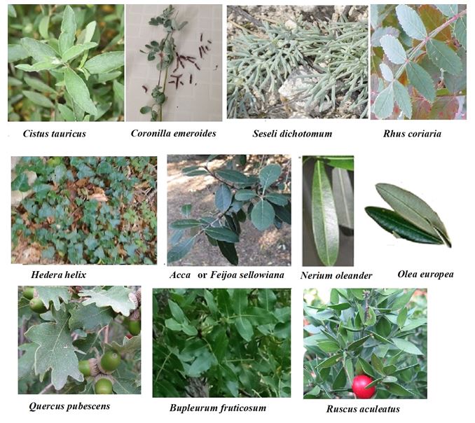 |
| Figure 1: The view of the plant surface belonging studied species. The blue color is marked |
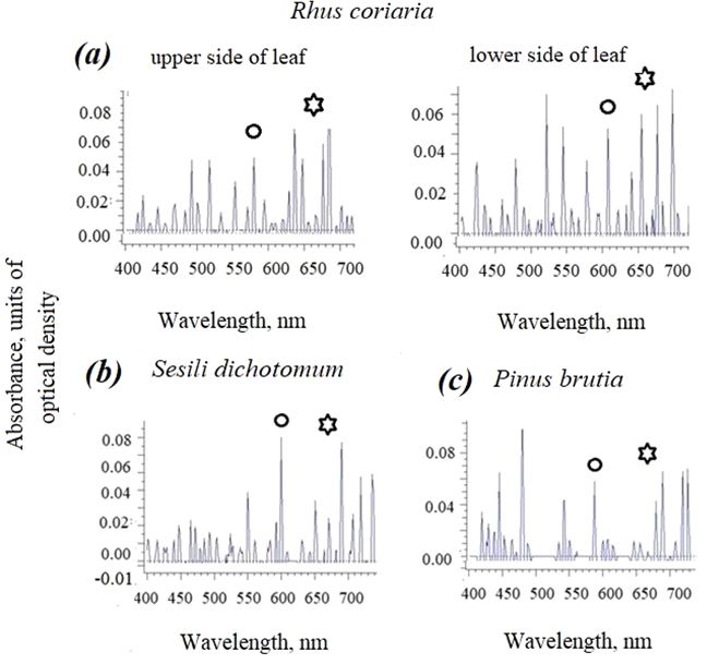 |
| Figure 2: The absorbance spectra of the intact surface of wide (a) and narrow (b, c) leaves of plants (a) Rhus coriaria; (b) Seseli dichotomum; (c) Pinus brutia. The maxima of azulenes are marked with circles, and the maximum of chlorophyll- with asterisks. |
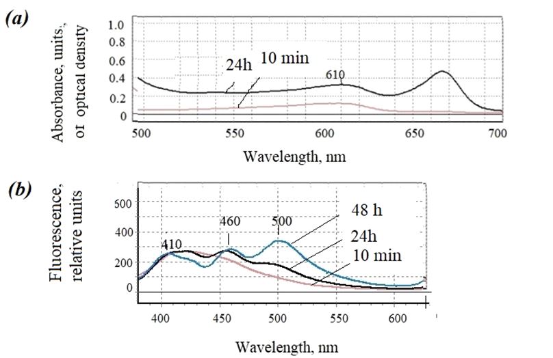 |
| Figure 3: Examples of absorption and fluorescence spectra of ethanol extracts -from leaves of Olea europea (a) and (b) Bupleurum fruticosum |
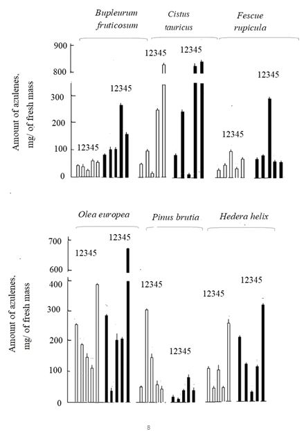 |
| Figure 4: The amount of azulenes in the ethanol extracts for 10 minutes in February (1), March (2), April (3), May (4), June (5). White columns based on fresh weight (weight), and black columns based on dry weight. |
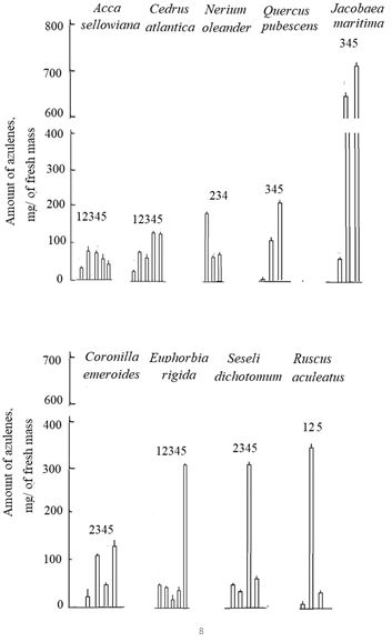 |
| Figure 5: The amount of azulenes in the 10 min ethanol extracts in February (1), March (2), April (3), May (4), June (5). Calculation for fresh weight. |
Introduction
Observations in natural conditions have shown earlier [1,2] that the blue or silver color of the leaves appeared to be due to the presence of blue pigments azulenes in some species. This event hypothethically may be applicated to pharmacy that made similar objects as potential resources of new medicinal azulenic drugs. We started to work in the problem by spectral methods, including microspectrophotometry [3-6]. Previously, the isolation and determination of azulenes was carried out mainly by distillation of essential oils, which took a lot of time and required a large amount of vegetable raw materials as well as usual analytic chemical methods. The importance of the search of new azulene-containing plants was also actual because assortment of similar species used mainly in folk medicine and cosmetics is narrow yet [7-9].
Main mechanism of the azulenes’ action is considered as antioxidant effects in medicine due to many unsaturated bonds in the main molecules [8,9]. Similar mechanism is proposed for plant cells themselves [3-6]. The surface of plant cells is sensitive to changes in ultraviolet radiation, tropospheric ozone concentration, salinity, traumatic factors, etc. that induced the formation of damaging reactive oxygen species. As a protection, plants secrete various compounds, in particular azulenes that are deposited on the surface. It has been suggested that azulenes may be involved in protecting the cell surface from damaging effects [4-6].
Primary experiments in the problem have been done in the subtropical zone of Russia, mainly on Caucasus [1,2]. However, these compounds have also been found in some species of the temporary climate zone [3]. In this case, it was needed to estimate perspectives of the species to synthesize and accumulate azulenes in a dependence on the season in different climatic zones.
The blue or silver color of the leaves appeared to be either constant or noticeable only in certain season. This is the question that interested us for the dry subtropics of the Crimea, where, compared with the region of the Russian temporary climate region, the highest ultraviolet insolation. There are many medicinal species in the dry subtropics of Crimea [10], but the inclusion of azulenes in their composition has not yet been described. Since this problem is important both for the ecological monitoring of these compounds and for the analysis of the resources of natural sources of azulenes with medicinal properties [7-9], the task was set to find out how the color appears and changes due to the presence of these blue pigments using spectral methods of their assessment [2,4].
Methods and Materials
Objects: The species of wild plants and introduced plants growing in Crimea with blue or silver leaf color were selected as the objects of research.
Places where Plants are Collected
Plant samples were taken in natural and anthropogenic uncontaminated territories of Crimea as a biosphere model. The plants were collected on the Southern coast of Crimea in the vicinity of the Nikitsky Botanical Garden (Yalta): wild species are found in natural forest high-juniper-downy-oak phytocenoses dominated by Juniperus excelsa M. Bieb. and Quercus pubescensWilld. And participation in the undergrowth of Cistus tauricus J. Presl. et C. Presl.; introductions – in parks and cultural plantings (gardens). Four more species were collected in coastal ecotopes of the western coast of Crimea (Saki – Yevpatoria) in the coastal saline ecotopes of the Black Sea (Table 1). For the purity of the experiment, leaf samples were collected once a month from the same plants or in the same population and in the same places.
Spectral Measurements of Intact Leaves
Absorption (absorbance) of the intact leaves was measured directly on slides using the microspectrophotometer/microspectrofluorimeter MSF-15 (LOMO, St. Petersburg, Russia) [8-10].The position of the maxima in the absorption spectra of intact cell surfaces was determined according to the Zolotarev method by the option of the reflection spectra differentiation [11]. For every species, the absorbance spectra were registered in 10 leaves (n= 10 measurements).
Spectral Measurements of Leaf Extracts
The absorption and fluorescence spectra of extracts with 100% acetone or 95% ethanol from cells (1:10 w/v for 10 min to 1 hour or more) in 1-0.5 cm cuvettes were recorded using the Unicam Helios-epsilon spectrophotometer (USA), spectrophotometer Specord M-40 (Germany) and Perkin Elmer 350 MPF-44 B spectrofluorometer (Great Britain) .The concentration of azulenes (A) was estimated in the ethanol or acetone extracts according to the [7,8].The results were expressed as an average value of ±SEM. For each variant, the average error of the experiment was calculated with three to four repetitions. The relative standard deviation was 5-6% (n=3-4 samples per species), P=0.95.
Results and Discussion
The surface of Crimean species was studied using by spectroscopic methods, including microspectrophorimetry and microspectrofluorimetry, obtaining their photographic images, the absorption and fluorescence spectra of the leaf intact extracts.
Figure 1 show that samples of plant leaf samples were taken from species with blue or silver colored surfaces. These samples had a bluish and blue color, which suggests the presence of blue pigments – sesquiterpene lactones, such as azulenes. The color did not belong to anthocyanin, because it did not change with a change in pH when we acted with acid or an alkaline medium.
The Absorption Spectra of Intact Leaves
The absorption spectra of whole leaves were recorded using a microspectrophotometer/microspectrofluorimeter. In order to determine the maxima in reflection spectroscopy in the microspectrophotometer a differentiation option was used to identify maxima in the absorbance spectra according to Zolotarev method [11]. Examples of such a method are shown for samples of sumac Rhus coriaria, fork weed Sesili dichotomum and Pinus brutia (Figure 2). To identify maxima in the region of 550-670 nm, it was enough to provide information with positive values [2].On Figure 2 one can see differences in maxima 580 and 610 nm between upper and lower sides of Rhus coriaria leaves, relatively. In narrow leaves of Sesili dichotomum and Pinus brutia there are maxima 600 and 590 nm. The absorbance maxima found in the spectra of leaves in the region of 580-620 nm were observed in many species (Table 2).
In Table 2 there are maxima in the azulene region and their optical density in the studied species, depending on the season. This is especially important for pharmacology in order to identify the periods of the greatest accumulation of azulenes. Differences in optical density are visible in the maxima characteristic of azulenes between the upper and lower surface of the leaf and in a dependence on the season. The highest values of optical density are at the upper side of Acca selwiana leaf in June, in April, as well as at the lower side of the leaf in Bupleurum fruticosum (May), Juniperus excelsa (March and June), Coronilla varia (April) and Seseli dichotomum (May).
In April, a number of plants, the optical density decreases in comparison with February and March (Helix, Jacobea, Juniperus, Olea, Quercus pubescens) or is not registered at all in the azulene region, as in Bupleurum. The highest values of optical density in the region of 580-620 nm were noted in May-June for Bupleurum, Seseli, Fescue, Juniperus, Jacobea, and Olea. But there is another variant of events – a decrease in optical density in June in the Seseli and Cistus. We saw two periods when the optical density is high – this is February-March for Bupleurum, Euphorbia, Seseli, Fescue, Juniperus, or after a decrease in April, it rises again in May-June, with the exception of the needle. There are very noticeable differences by season, especially in Bupleurum, Cistus and Fescue. Bupleurum has the most intense azulene season in February, a failure in March and beyond increase to June. The choose of a pleasant time for the collection of raw pharmaceutical material in maximal amount during all spring- June should be for azulene-enriched species.
The Absorbance and Fluorescence Spectra of Extracts
To confirm the presence of azulenes on the surface of the leaves of the studied plants, experiments were further conducted on the extraction of pigments and the study of their spectral composition to find out whether the data obtained on the detection of azulenes by microspectrophotometry were consistent with them.
Two fractions of extracts were analyzed – 1. infusing the leaves for 10 minutes, where the outer layer of azulenes is washed off the surface [1-3] and prolonged infusing for 24 or 48 hours, when intracellular azulenes can be extracted [4,5]. Thus, it is possible to compare the content of azulenes on the outside and inside the cell. In Figure 3 the results of a 10-minute extraction are shown, where blue pigments with maxima in.
The regions of 580-620 nm give the extracts the highs characteristic of azulenes. In a short time from 5-10 minutes, they are mostly washed off the outside of the leaf, still weakly penetrating inside and not yet extracting chlorophyll from chloroplasts. If the wax layer or cuticle features allow this to be done, as shown earlier [1,2]. Maxima in the azulene region are visible in all studied species. From the data in Figure 3., the largest maximum of 610 nm in olives in the absorption spectrum of 24-hour extracts, and the presence of chlorophyll with a maximum of 666 nm is visible. According to such absorption spectra at the maximum characteristic of azulenes, their content in the ethanol extract is analyzed. The fluorescence spectra of extracts can also show the peaks of 410, 460, and 430 nm characteristic of azulenes, as shown for Bupleurum (Figure 3), visible both at 10 minutes and at 24 hours
The Azulenes’ Content
We analyzed the content of azulenes in leaf extracts depending on the month of harvest. Based on the obtained absorption spectra of extracts, the azulene content was determined every month from February to June, and in a number of species later (July-September). The data obtained can be conditionally divided into three main trends, starting from the initial date of observation; 1. An increase in the content of azulenes; 2. Their decrease; 3. Their approximately stable concentration.
Figure 4 shows the data based on the raw and dry weight of the leaves. As experiments have shown, there are no significant differences in the percentage ratio, therefore, in the following Figure 5, we provide only data on raw weight. 10 minute extracts were analyzed in detail, which showed the most noticeable changes (Figures 4 and 5). Based on the obtained absorption spectra of the extracts, the azulene content was determined every month from February to June.
On Figure 4, the increase in the concentration of azulenes on the side of the leaves of Cistis is more clearly visible, to a lesser extent in Fescue and Bupleurum. Sometimes, the increase is single and sudden, for example, in June, as in olives and ivy, or in March as for the Pinus brutia.
There is a sudden increase in azulenes by May-June in Euphorbia, Cedrus anlantica, Cistis, Quercus, Jacobea (Fig.), but in March this is also noticed niglice. A significant decrease in blue pigments on the side of the leaves was found in olive and oleander, compared with the beginning of the observation.
All of the above refers to the relative concentration of azulenes, and we associated it with the protective function in the spring and summer period from damage by high ultraviolet radiation and ozone.
Along with a 10-minute extraction, seasonal data on the infusion of leaves in alcohol for 24 hours were obtained. Prolonged extraction of blue pigments for 24 hours with these organic solvents indicates that azulenes are also present inside the cells. This great material will be published in a special article. In this work, for a number of plants especially enriched with blue pigments, we compared in Table 3 the concentrations of azulenes in 10 minute extracts (on the side of the cells) and 24 hour extracts (inside the cells).
It should be noted that the concentration of azulenes inside the cells, compared with the concentration on the surface in February, is about five times higher in Bupleurum, and more than 2 times in frankincense and olives. But in May, in the first type, the difference between 10-minute and 24-hour extracts becomes smaller in Bupleurum, almost equaled Cistis, and increased up to 7 times inside the olive cells. If we compare the data in February and May, we see a slight increase in azulenes on the opposite side in Bupleurum, a significant (up to 5 times) in Cistis, but a twofold decrease in olives. On the contrary, inside the cells of Bupleurum and Olea, the differences in February-May are small, whereas in Cistis the concentration of these pigments doubled in spring. Thus, the content of azulenes inside cells also varies significantly over the months of collection, although differently compared to the frequency.
The question arises, what role do azulenes play inside cells? It was also found that the redox properties of artificially synthesized azulene allow azulenes to be electron donors in the photosynthetic electron transport chain of photosynthetic membranes [2,5]. This is important in case of damage to chloroplasts, and thus, again, we can talk about the protective role of azulenes in this case. Moreover, the blue pigments were also found in isolated chloroplasts of three clower species [12].
Unlike plants that are actively developing in spring and summer, at the end of the summer season (in July- early September), noticeable differences in the content of azulenes can also be observed in species growing in arid conditions, as can be seen from the data in Table 4. In Crambe, which grows on coastal sands and pebbles in the coastal zone of the Black Sea, during the study period there was a doubling of the number of azulenes on the cell side (10 minute extract), and a comparative small change in the concentration of these pigments inside the cells from July to early September (24 hour extracts). A similar pattern was noted in the blue-headed Eryngium. But in Eleagnum and Leymus, the number of lateral azulenes did not change much. But inside the cell, the concentration of these pigments increased almost twice, and in the grate it decreased almost four times. Apparently, differences in azulene metabolism in watered or arid areas are also associated with the protective function of azulenes.
Looking through the data, it is possible to identify several species whose leaves are enriched with azulenes. First of all, this is olive Olea, oleaster Elaeagnus angustifolia, relict rock rose Cistus tauricus among woody plants or among wild herbs - Euphorbia rigida, Seseli dichotomum and Jacobaea maritima. Previously, they were not noted among medicinal plants including azulenes [10]. In this regard, screening of azulene-containing plants in Nature by spectral methods may be of interest not only for environmental monitoring, but also as a definite resource for pharmacology. Unfortunately, since the 90s of the twentieth century, studies of natural azulenes have not been conducted. With the help of mass spectrometry, it was possible to determine the chemical nature of a few species, and this will be the subject of further research.
Among plants studied, May- June more optimal for the herbs collection like Bupleurum fruticosum, Cistis tauricus, Cistus tauricus and Jacobea maritima (Sinecio cineraria) enriched in azulenes. Second and third species are known as medicinal ones [10,13], as well as many plants of genus Bupleurum, although for B. maritima it is unknown yet. The above-mentioned plants possess spasmolitic and anticeptic features [13]. It is possible to include azulenes as active drug matter for their species.
Conclusion
The use of spectral methods made it possible to obtain information about the presence of azulenes on the surface and inside the leaves of plants growing in the Crimea. In a significant number of plants with blue or silver color, the absorption spectra showed maxima of 580-620 nm, characteristic of blue azulene pigments, which is confirmed by their appearance in 10-minute and 24-hour extracts with ethanol or acetone. It is likely that azulenes have a protective function due to their antioxidant properties. The data obtained show that the accumulation of azulenes on the cell surface, possibly in the cuticle, occurs in April-May, when the highest ultraviolet insolation and the maximum formation of ground-level ozone. Then the appearance and accumulation of azulenes can be considered as a protection against the formation of reactive oxygen species.
Contribution of the Authors
The idea of the work, all experiments, writing the article (Roshchina V.V.), identification of plants, collection of material and editing of the article (Kraynyuk E.S.),
Conflict of Interest
Authors have no conflict of interest.
Use of AI Tools Declaration
The authors declare they have not used Artificial Intelligence (AI) tools in the creation of this article.
Ethical Approval
The author declares that the study was carried out following scientific ethics and conduct. This study did not involve any use of animals, hence no ethical approval has been obtained from the concerned committee. Authors had no grants for the experiments and writing of the paper.
Acknowledgments
Authors are thankful to the Optical Microscopy and Spectrophotometry core facilities, ICB RAS, Federal Research Center “Pushchino Scientific Center for Biological Research of the Russian Academy of Sciences” for possibility to work in optic department and especially to engineer Nadezhda K. Prizova and technical assistant Lubov’ M. Khaibulaeva.
References
- Roshchina VV, Kuchin AV, Kunyev AR, Soltani GA, Khaibulaeva LM, Prizova NK (2022) The presence of azulene on the surface of plant cells as a test for ozone sensitivity. Biochemistry (Moscow), Supplement ser A: Membrane and Cell (Biological Membranes). 16: 167-74.
- Roshchina VV, Yashin VA, Kynyev AR (2023) Study of the Spectral Characteristics of the Plant Cell Surface: Occurrence of Azulenes and Biogenic Amines. , Biochemistry (Moscow), Supplement Series A: Membrane and Cell Biology, 17: 276-85.
- Roshchina VV, Prizova NK, Khaibulaeva LM (2022) Azulenes of the leaf surface as a protective optical filter. Russian journal of biological physics and chemistry, 7: 36-9.
- Roshchina VV, Kunyev AR, Fateryga VV, Shovkun MM (2023) Application of microspectrofluorimeter/microspectrophotometer for the study of the surface of plant cells. Russian Journal of Biologica Physics and Chemistry, 8: 137-42.
- Roshchina VV (2023) Plant leaf surface as a sensory system in allelopathic relations: 1. Role of azulenes. Allelopathy Journal. 59: 109-22.
- Roshchina VV (2022) Possible role of azulenes in plant life: Experiments with Models. SMP Environ Sci Technol, 1: 1-10
- Konovalov DA (1995) Natural azulenes. Biological Resources (Biologicheskie Resursy, in Russian) 31: 101-30
- Bakun P, Czarczynska-Goslinska B, Goslinski T, Lijewski S (2021) In vitro and in vivo biological activities of azulene derivatives with potential applications in medicine. Med Chem Res. 30: 834-46.
- Murfin LC, Lewis SE (2021) Azulene- a Bright core fore sensing and imaging. Molecules, 26: 353-62.
- Krainyuk ES (2018) Medicinal Plants of Crimea. Illustrated book. Business-Inform: Simferopol, 512 pp.
- Zolotarev VM (2012) Application of differentiation in reflection spectroscopy. Optics and Spectroscopy, 112: 150-4
- Roshchina VV (2024) Azulenes in Plant Cell: Clover as their Useful Resource. Annals of Agricultural &Crop Sciences, 9: 1147
- Golovkin BN, Rudenskaya RN, TrofimovaI A, Shreter AI (2001) Biological Active Substances of Plant Origin. M: Nauka, in three volumes, 350 + 764 + 216 pp.
Article Information
Research Article
Received Date: January 01, 2025
Accepted Date: February 01, 2025
Published Date: February 04, 2025
Spectral Studies for the Search for Azulene-Containing Plants in Dry Subtropics of the Crimea
Volume 4 | Issue 1
Citation
V.V. Roshchina, E.S. Kraynyuk (2025) Spectral Studies for the Search for Azulene-Containing Plants in Dry Subtropics of the Crimea. J Plant Biol Agron 4: 1-14
Copyright
©2025 V.V. Roshchina. This is an open-access article distributed under the terms of the Creative Commons Attribution License, which permits unrestricted use, distribution, and reproduction in any medium, provided the original author and source are credited.

doi: jpba.2025.4.101