

Review Article
Volume-1 Issue-1, 2025
A Giant Pediculated Vulvar Lesion: A Case Report
-
Received Date: April 02, 2025
-
Accepted Date: April 21, 2025
-
Published Date: April 28, 2025
Journal Information
Abstract
Angiofibroma is a benign mesenchymal tumor. It’s clinical presentation is nonspecific and similar to other vulvar tumors. Angiofibroma has good prognosis,despite some risk of recurrence.We present a 44-year-old woman with a bulky left vulvar lesion.
Key words
Angiofibroma; Mesenchymal; Tumor; Vulva
 |
 |
Introduction
Angiofibroma is a soft tissue tumor that occurs in vulvo vaginal region in females and inguino-scrotal region in males [1].
These tumors are well-circumscribed,superficial and includes soft spindle cells and veins [2]. It is first described by Nucci et al.in 1997 in a series of 6 cases that occurred almost in the vulvar region of middle-aged women [3].
Since the first description by Nucci, few studies have been published about this kind of lesion in the literature. According to studies, there is no known risk factor for angiofibroma.
Case Presentation
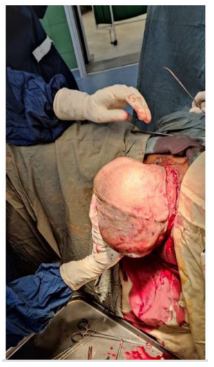
We present an unusual case of pediculated angiofibroma mass. A healthy virgin 44-year-old woman consulted for a painless vulvar mass. There was no previous infection or bleeding. The patient history revealed that the mass was first identified four years earlier and it had gradually enlarged. It caused discomfort when sitting or walking due to its size. Gross examination showed a firm freely mobile tumor meassuring 25*20*17 cm in the left labia majora.
There was no family history of cancer. She didn’t report any use of OCP or hormone replacement therapy.The patient gynecological examination was normal, and no palpable inguinal lymph nodes were distinguished.
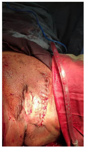
The patient underwent surgery with a diagnosis of vulvar mass. The mass was composed of large vessels and after resection had 3 kgs weight. Microscopic evaluation showed a vulvar tissue with a well defined neoplastic growth composed of fassicle of spindle cells with bland nuclei along with thickwall dilated blood vessels.
Discussion
Angiofibroma is considered benign.Surgical excision of the lesion seems to be the adequate management [4], not only to treat the patient but also to achieve a correct diagnosis.The potential recurrence risk of these lesions is low [5].
However, Mc Cluggage et al. reported one case of recurrence in a 49-year-old woman that occurred 6 months after excision [6]. Angiofibroma should be considered in the differential diagnosis of painless soft masses that may reach large dimensions in the vulva [7].
The present case is the largest angiofibroma defined in the literature.
There are many mesenchymal tumors which enter into the differential diagnosis with angiofibroma as spindle cell lipoma, solitary fibrous tumor, mammary-type myofibroblastoma, angiomyofibroblastoma, aggressive angiomyxoma and smooth muscle tumor. All these described tumors share similar morphologic features and are characterized by bland ovoid to spindle- shaped cells with wispy collagen, vaiably sized thichwalled blood vessels [8].
Conclusion
In summary, Pathologists should be aware of morphological variation to avoid diagnostic errors and therefore an aggressive treatment. Angiofibroma in women represents a benign lesion , so a treatment of simple local excision of the lesion is adequate.
References
- MR Nucci, SR Granter, CDM Fletcher (1997) Cellular angiofibroma: a benign neoplasm distinct from angiomyofibroblastoma and spindle cell lipoma,” American Journal of Surgical Pathology 21: 636-44.
- E Chen, CDM Fletcher (2010) Cellular angiofibroma with Atypia or sarcomatous transformation:clinicopathologic analysis of 13 cases American J Surgical Pathology 34: 707-14.
- Nucci MR, Granter SR, Fletcher CDM.Cellular angiofibroma:a benign neoplasm distinct from angiomyofibroblastoma and spindle cell lipoma.Am J Surg Pathol 21: 636-44.
- Kumar P, Singh S, Kumar A, Janoria S (2018) A rare case of cellular angiofibroma affecting the periurethral region in a 38-year-old woman.BMJ Case Rep
- Khmou M, Lamalmi N, Malihy A, Rouas L, Alhamany Z (2016) Cellular angiofibroma of the vulva: a poorly known entity, a case report and literature review. BMC Clin Pathol 16:8.
- Mc Cluggage WG, Perenyei M, Irwin ST (2002) Recurrent cellular angiofibroma of the vulva. J Clin Pathol 55: 477-9
- Umit A, Hasan T, Unal T, Ahmet TE, Ahmet K (2016) A Giant Vulvar Mass: A Case Study of Cellular Angiofibroma.Hindawi Case Reports in Obstetrics and Gynecology2016: 2094818.
- Micheletti AM. Silva AC, Nascimento AG, Da Silva CS, Murta EF, et al. (2005) Cellular angiofibroma of the vulva:case report with clinicopathological and immunohistochemistry study. Sao Paulo Med J 123: 250-2.
Artcle Information
Review Article
Received Date: April 02, 2025
Accepted Date: April 21, 2025
Published Date: April 28, 2025
European Journal of Case Reports
Volume 1 | Issue 1
Citation
ELaouni.Soukaina (2025) Rapid Growing Amyloid Goiter Mimicking a Malignant Thyroid Tumor in 24-Year-OldMale, Secondary Amyloidosis and End-Stage Kidney Failure. Eur J Case Rep 1: 103
Copyright
©2025 ELaouni.Soukaina. This is an open-access article distributed under the terms of the Creative Commons Attribution License, which permits unrestricted use, distribution, and reproduction in any medium, provided the original author and source are credited.

doi: ejcr.2025.1.103