

Review Article
Volume-1 Issue-1, 2025
The Glaring Zebra: A Case of Hypothyroidism-associated Recurrent Massive Pleural Effusion
Received Date: July 04, 2025
Accepted Date: July 22, 2025
Published Date: July 29, 2025
Journal Information
Abstract
Introduction
Pleural effusions can be secondary to a myriad of conditions. Rarely can it be due to hypothyroidism as well. We present a case of massive pleural effusion resulting from hypothyroidism.
Case
A 75-year-old male with a history of liver cirrhosis, hypothyroidism and medication non-adherence was brought into the emergency department (ED) with shortness of breath and altered mental status. Physical exam and chest imaging was consistent with right sided pleural effusion. Effusion was exudative in nature. The hospitalization was complicated by multiple recurrences despite thoracentesis and pleurodesis. Labs revealed hypothyroidism and finally the patient was started on hormone replacement resulting in resolution of the effusion.
Discussion
Pleural effusion is a rare manifestation of hypothyroidism which is thought to be mediated by vascular endothelial factor. Both exudative and transudative patterns can be seen on pleural fluid analysis. Mainstay of treatment is hormonal replacement.
Conclusion
The clinicians need to be cognizant of the rare etiologies of pleural effusion. Due work-up should be done depending on patient presentation for a timely diagnosis and management.
Key words
Hypothyroidism; Pleural Effusion; Thyroid Hormone; Endocrine Disorder; Myxedema
Abbreviations
PEF- Pleural Effusion; ULN- Upper Limit of Normal; TSH- Thyroid Stimulating Hormone; FT4- Free T4; VEGF- Vascular Endothelial Growth Factor; CT- Computer Tomography
Thyroid Function Test |
5/10/2023 (admission) |
th |
6/1/2023 |
TSH (uIU/m) |
323.6 |
101.7 |
15.3 |
FT4 (ng/dL) |
0.29 |
0.49 |
0.90 |
Fluid Analysis |
5/10/2023 |
5/12/2023 (2nd day) |
5/30/2023 |
Pleural fluid LDH/serum LDH |
146/279 (> 2/3 ULN of S. LDH) |
144/227 (> 0.6) |
507/147 (> 0.6) |
Pleural fluid protein/serum protein |
4.9/6.8 (>0.5) |
4.5/5.7 (>0.5) |
3.9/6.2 (>0.5) |
 |
 |
Case Report
Introduction
Hypothyroidism is a systemic endocrine disorder resulting from inadequate synthesis, secretion, or biological effects of thyroxine, leading to myriad non-specific clinical presentations [1]. Pleural effusion (PEF), defined as the accumulation of fluid in the pleural space between the parietal and the visceral pleura due to an imbalance between fluid formation and removal [2,3], is an uncommon manifestation of hypothyroidism. Massive pleural effusion from hypothyroidism is particularly very uncommon [4]. Some of the more common causes of PEFs include congestive heart failure, nephrotic syndrome, malignancy, and pneumonia [4]. Herein, we present a patient with hypothyroidism-induced pleural effusion, providing insight into the clinical features, diagnostic workup, management, and outcome.
Case Presentation
A 75-year-old Caucasian male with a history of liver cirrhosis, chronic kidney disease, hypothyroidism, and medication non-adherence was brought into the emergency department (ED) with progressively worsening shortness of breath and altered mental status via the emergency medical service. Reportedly the patient was having some shortness of breath for the past few days which was gradually progressive. He had some associated right sided chest discomfort. On the day of presentation, he was reported to be confused and altered. He had no preceding head trauma, fever, cough, focal weakness, orthopnea, paroxysmal nocturnal dyspnea, or recent long-distance travel.
Vitals signs revealed a temperature of 91.9 F, pulse rate of 53 bpm, BP 156/106 mmHg, and SPO2 of 89% on room air. On the examination, the patient was in obvious respiratory distress, lethargic, and oriented only to person. Chest examination revealed markedly diminished breath sounds and dullness to percussion over the right lung base. The extremity exam revealed bilateral non-pitting pedal edema. The remainder of the examination was unremarkable.
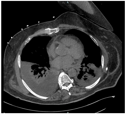
Investigation revealed a white blood cell count (WBC) of 6.0 x 10 /mm3, hemoglobin of 12.9 g/dL, and creatinine of 3.25 mg/dL (baseline of 1.4-1.9). Liver function and lactate were within normal limits. Chest X-ray demonstrated severe rightsided pleural effusion with complete opacification of right hemithorax (figure 1), prompting further evaluation. A chest CT scan performed on 5/12/2023 (2nd day of admission), showed a large right and small left pleural effusion with lower lobe atelectasis and mild ground-glass opacities in right upper lobe without any associated adenopathy (figure 2).
The patient underwent thoracentesis with pleural fluid analysis showing exudative physiology which was inconsistent with hepatic hydrothorax in the setting of known liver cirrhosis (Table 1). Other laboratory investigations, including pleural fluid cell count, glucose, pH, and cytology, were unremarkable. Given the persistence of the altered mental status, thyroid function test and serum cortisol were checked which revealed a TSH of 323.5 uIU/mL, however, the cortisol was unremarkable. The patient's home dose of oral levothyroxine was continued. The hospital course was complicated by re-accumulation of exudative pleural fluid requiring multiple thoracentesis to relieve dyspnea. A chest tube was eventually placed. Having ruled out more common etiologies of exudative effusion including infection and malignancy, severe untreated hypothyroidism was pursued as the likely etiology for pleural effusion. The patient was switched to intravenous levothyroxine given the overall patient presentation and concern for myxedema coma. The patient remained on the treatment and ultimately levothyroxine was later switched to oral route after 8 days of treatment as the patient’s mentation had improved. Repeat TSH was 101.7 uIU/mL. The patient remained on oral levothyroxine and the patient’s labs showed an improved TSH of 15 uIU/mL with free T4 of 0.90 ng/dL at the time of discharge, with significant improvement in mental status. A repeat chest X-ray revealed the resolution of pleural effusion. The patient was discharged on oral levothyroxine with outpatient follow-up.
Discussion
Hypothyroidism is a very ubiquitous disease with nonspecific clinical manifestations including easy fatigability, dry and coarse skin, cold extremities, decreased appetite, hoarse voice, weight gain, depression, and psychomotor retardation [5]. Mild serous effusion such as ascites, pericardial, and pleural effusions are not uncommonly seen but are typically asymptomatic [6]. In rare cases, massive pleural effusion presenting with respiratory compromise may be the primary presentation [7]. Owing to the nonspecific presentation, diagnosis is usually biochemical and is characterized by elevated serum TSH levels.
The incidence of pleural effusion (PEF) in hypothyroidism has been estimated at 10 to 30%, associated with small fluid accumulations, and of limited clinical significance [8]. However, this may be overestimated as most of the pleural effusion in hypothyroidism is related to comorbid conditions [9]. The exact incidence of hypothyroidism-induced PEF remains unclear [9]. Massive PEF has been reported as an exceedingly rare yet serious complication of decompensated hypothyroidism which is under recognized [9]. This is thought to be due to longstanding untreated hypothyroidism [8]. Studies have reported a mortality rate of approximately 60%, making an early diagnosis crucial for a favorable clinical outcome[10].
The pathophysiology of hypothyroidism-induced PEF has been postulated to be due to increased systemic capillary membrane permeability mediated by vascular endothelial growth factor (VEGF) and disruption in electrolyte metabolism related to hypothyroidism [4,10]. The resultant extravasation of albumin and disturbances in lymphatic drainage leads to exudative physiology being commonly demonstrated on pleural fluid analysis in cases of hypothyroidism-induced PEFs [4]. Although initially thought to be related to autoimmune-related serositis seen commonly in concomitant autoimmune disorders associated with Hashimoto’s thyroiditis, this hypothesis does not seem to explain the occurrence of pleural effusion in iodine deficiency-related hypothyroidism [7]. Significantly, sometimes the effusions can be border line between exudative and transudative with no evidence of inflammation [9]. There have also been reports of unilateral and bilateral cases of hypothyroidism-induced pleural effusion [10]. Ultimately, these variations pose a challenge to the diagnosis of hypothyroidism-related pleural effusion.
In our patient, we initially made a diagnosis of pleural effusion with differentials of parapneumonic effusion, TB, hepatic hydrothorax, heart failure, and malignant effusion. Fluid analysis was exudative making heart failure and hepatic hydrothorax less likely, especially so given the patient had evidence of well-compensated liver function. There was concern for parapneumonic effusion from possible aspiration given the patient had altered mental status, but the fluid analysis revealed no evidence of infection. Fluid cytology was also negative for malignant cells. However, due to the low sensitivity of the test; we repeated thoracentesis, but cytology results remained negative [10]. Considering the patient had persistent altered mental status with psychomotor retardation and bilateral non-pitting pedal edema, the TSH was checked which was markedly elevated with low free T4 and T3 levels. A review of the literature revealed rare cases of pleural effusion in hypothyroidism which were treated with timely hormonal replacement therapy [11]. Our patient had already been on oral levothyroxine since admission. We switched him to intravenous therapy in light of suspicion for possible myxedema coma contributing to altered mentation and pleural effusion. The American Thyroid Association recommends an intravenous levothyroxine dose of 75% of the oral dose. Intravenous injection is strongly not recommended for long-term treatment due to its rapid and great effect on thyroid hormone homeostasis [12].Hence, intravenous treatment was pursued for 3 days only. The patient was monitored closely, and his thyroid function improved on the therapy. His mentation also improved, and no further pleural effusions were noted. The patient was later switched back to oral levothyroxine therapy.
Although pleural effusion is commonly seen in hypothyroidism, they are rarely caused by hypothyroidism. Even more rarely do they become clinically significant; thus, posing diagnostic challenges. Nonetheless, other competing diagnoses, including infectious, neoplastic, autoimmune and metabolic diseases, must be ruled out first. Given the complete resolution of the pleural effusion after hormonal replacement with no recurrence after achieving a euthyroid state, it suggests that the pleural effusion was likely related to hypothyroidism.
Conclusion
This case report highlights the association between hypothyroidism and pleural effusion, emphasizing the importance of considering thyroid dysfunction in patients presenting with unexplained pleural effusions. Early diagnosis and treatment of hypothyroidism is essential to prevent further complications and improve patient outcomes. Further research is warranted to elucidate the exact mechanisms underlying this association and identify potential therapeutic interventions.
Acknowledgement
Piedmont Athens Regional Medical Center
References
- Yuan GQ, Yan QL, He MH (2020) A Case of Pleural Effusion Caused by Hypothyroidism. Journal of Biosciences and Medicines, 8: 1-4
- Saguil A, Wyrick K, Hallgren J (2014) Diagnostic approach to pleural effusion. Am Fam Physician, 90: 99-104.
- Karkhanis VS, Joshi JM (2012) Pleural effusion: diagnosis, treatment, and management. Open Access Emerg Med, 4: 31-52
- Lee JH, Park M, Park MJ, Jo YS (2018) Massive pleural and pericardial effusion due to hypothyroidism in a patient with a surgically treated thyroid-stimulating hormone-producing pituitary adenoma. Acta Clin Belg, 73: 398-401
- Yuan GQ, Yan QL, He MH (2020) A Case of Pleural Effusion Caused by Hypothyroidism. Journal of Biosciences and Medicines, 8: 1-4
- Guha B, Krishnaswamy G, Peiris A (2002) The diagnosis and management of hypothyroidism. South Med J, 95: 475-80.
- Sachdev Y, Hall R (1975) Effusions into body cavities in hypothyroidism. Lancet, 1: 564-6.
- Gomes Santos P, Calças Marques R, Martins Dos Santos P, Carreira da Costa C, Mogildea M (2023) Ascites, Pleural, and Pericardial Effusion in Primary Hypothyroidism: A Rare Case Report. Cureus, 15: e50429.
- Rehan M, Alam MT, Imran K, Farrukh SZUI, Masroor M, Kumar P (2013) The frequency of various diseases in patients presenting with pleural effusion. Gomal Journal of Medical Sciences, 11.
- Gottehrer A, Roa J, Stanford GG, Chernow B, Sahn SA (1990) Hypothyroidism and pleural effusions. Chest, 98: 1130-2.
- Pairman L, Beckert LEL, Dagger M, Maze MJ (2022) Evaluation of pleural fluid cytology for the diagnosis of malignant pleural effusion: a retrospective cohort study. Intern Med J, 52: 1154-9.
- Liu H, Li W, Zhang W, Sun S, Chen C (2023) Levothyroxine: Conventional and Novel Drug Delivery Formulations. Endocr Rev, 44: 393-416.
Artcle Information
Review Article
Received Date: July 04, 2025
Accepted Date: July 22, 2025
Published Date: July 29, 2025
European Journal of Case Reports
Volume 1 | Issue 1
Citation
Fawwad Ansari, Sammudeen Ibrahim, Anis Abobaker, Mubashira Aftab, Christopher March (2025) The Glaring Zebra: A Case of Hypothyroidism-associated Recurrent Massive Pleural Effusion. Eur J Case Rep 1: 105
Copyright
©2025 Fawwad Ansari. This is an open-access article distributed under the terms of the Creative Commons Attribution License, which permits unrestricted use, distribution, and reproduction in any medium, provided the original author and source are credited.

doi: ejcr.2025.1.105