

Review Article
Volume-1 Issue-1, 2021
Molecular Characterization of Kinetoplastida, Trypanosmatidae Isolates of Dogs and Cattles from Jos, Plateau State of Nigeria Using Polymerase Chain Reaction (PCR)
Received Date: August 20, 2022
Accepted Date: September 20, 2022
Published Date: September 23, 2022
Journal Information
Abstract
The study was designed to characterize trypanosomes isolates of dogs and Cattles from Jos, Plateau State of Nigeria to demonstrate presence of species and subspecies of different trypanosomes and its implications in these vertebrates. A total of 169 buffer coats of blood drawn from dogs (88) and Cattles (81) were examined by microscopy, five of these were positive for trypanosomes. The positive samples were characterized using Kin primers previously validated for differentiation of the following trypanosome stocks: Trypanosoma vivax (150bps, 250bps, 305bps); T.congolense (750bps and 780bps), T. b. brucei (540bps) and T. simiae (435bps). All positive samples from dogs showed 250 and 305 bps bands, that is, different forms of T. vivax while the third sample from dog additionally showed T. congolense (750 bps) band. The Cattle isolates showed 750bps and 500bps bands corresponding to T. congolense and T. b. brucei respectively. The trypanosomes infected Cattles and dogs showed an overall mean packed cell value (PCV) of 24.8 ± 26.6 (p< 0.0001) while noninfected animals PCV was 32.5 ± 40.6. The PCV results are consistent with anemia expected in trypanosomes infected animals. Thus, we characterized the presence of T. vivax and T. congolense in dogs, as well as T. congolense and T. brucei in Cattle and, from PCV, we demonstrated that the presence of these trypanosomatids resulted in in anemia for these animals.
Key words
PCR, Trypanosoma, Trypanosomiasis, Cattle,Dogs, Tsetse Fly
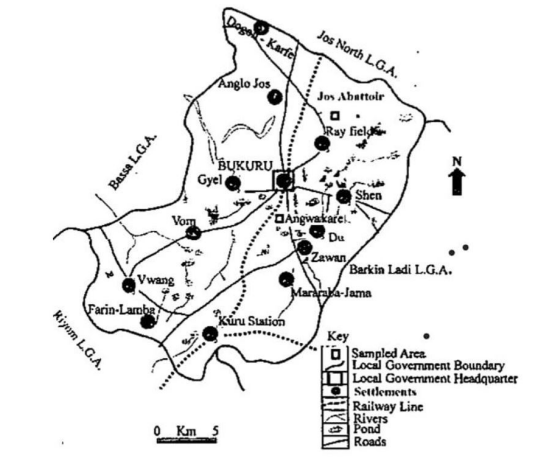 |
| Figure 1: Jos South Local Government Area of Plateau State |
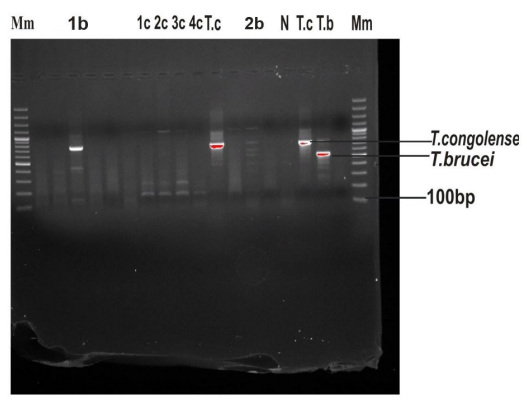 |
| Figure 2: Identification of the trypanosomes by PCR. Ethidium bromide stained gel showing PCR products (bovine samples:1b,2b) and (canine samples:1c,2c,3c,4c) with KIN 1 and KIN 2 primer sets. The Molecularweight marker are at the extreme lanes (Mm). Negative control (Lane N) was included while T.brucei (Lane T.b) and T.congolense (Lane T.c) were showed positive controls |
Introduction
African animal trypanosomiasis is a complex debilitating protozoan diseases of animals and humans [1,2]. It is transmitted mainly by tsetse fly limiting the use of large areas of land for livestock production [3]. Three elements are involved in transmission cycles, the mammalian reservoirs of the parasites, the tsetse fly that serves as cyclic vectors for transmission and the pathogenic parasite, the trypanosomes. African animal trypanosomiasis are ranked among the top ten Cattle diseases [4]. They affect all domestic animals. In West Africa, the major species of the trypanosomes that affect animals are Trypanosoma congolense, T. vivax, T. brucei and T.simiae whilst T. vivax is the most important agent for cattle infection in Brazil [5]. In tsetse region of Africa, T. b. brucei is probably the most important however, the most closely related to it, the T. evansi, is not transmitted by tsetse flies but mechanically by biting flies [6]. The T. brucei complex includes T.brucei brucei and the human T. b. gambiense and T. b. rhodensiense subspecies. While T. b. gambiense is found mostly in West Africa and causes chronic infection the T. b. rhodesiense causes fulminating infection and is resident in East and Southern Africa. Thus, understanding a whole interplay of transmission between animal reservoir and reemergence of both human and animal trypanosomiasis need to be kept in constant review.
For decades arguments have persisted as to whether some domestic animals including dogs are really reservoir hosts for trypanosomes or merely sentinels for trypanosomes infections since they succumb to infection in weeks after infection [7,8]. Authors like [9,10] have argued that variety of trypanosomes including T. congolense, T. evansi and T. brucei are hosts to canine trypanosomiasis. Dogs challenged by diverse species of trypanosomes show varying degrees of virulence and pathogenicity hence may act as source of infections to other domestic animals [11]. Recently, [12] have linked some domestic animals including dog and ruminants as potential reservoirs of human trypanosomiasis in Chad. Thus the study was designed to characterize trypanosomes isolates of dogs and Cattles from Jos, Plateau State of Nigeria, to demonstrate presence of species and subspecies oof different trypanosomes and its implications in these vetebrates.
Materials and Method
SiteA total of eighty-eight (88) blood samples were randomly collected from dogs at Angwankare, Bukuru and 81 blood samples from Cattles in Jos. Both areas are located in Jos South Local Government area of Plateau State. he area is about 1,250 metres above sea level.
Sample Collection and PreparationAnimals were randomly selected as described by and bled from jugular vein using sterile hypodermic needle into EDTAanti coagulant tubes [12]. A total of 5ml of blood sample was collected from each animal and properly labeled. The specimen bottles were gently rotated to ensure the blood samples were properly mixed with the anti-coagulant, and then kept in a cooler containing ice pack and immediately taken to the laboratory of The Veterinary Research Institute, Vom for processing.
Parasitological DiagnosisHematocrit centrifuge tubes (75 x 1.5mm) were filled with fresh blood containing anticoagulant (70µl). The tubes were sealed with clay and placed in a hematocrit centrifuge and centrifuged at 3000g for 5 minutes. The buffy coat appeared between the plasma and erythrocytes which was less than 1% of the total volume of the blood sample. Smears were prepared by scratching and breaking the capillary tubes 1mm below the surface of the buffy-coat and a drop of the buffy coat was expelled onto light microscope slide, smeared, and covered with a cover slip (18 x18 mm), then viewed using X40 objective lens. Approximately 200 fields of the preparations were examined for the presence of trypanosomes as described by [14]. The packed cell volume (PCV) percentages were read using a hematocrit reader.
Giemsa or Field’s-stained thin blood smear were also prepared and approximately 50 fields of the stained thin smears were examined with X100 oil immersion objective lens. Negative samples contained no trypanosomes. In the case of positive specimens, about 20 extra fields were examined to ascertain if more than one species of trypanosome were present as described in [15].
DNA ExtractionDNA was extracted from the buffy coat using DNA Mini kit (Qiagen,Germany) as described by the manufacturer. Briefly Qiagen protease (20 µl) were pipetted into the bottom of 1.5ml micro centrifuge tubes to which buffy coat (200 µl) and 200 µl of lysis buffer (AL) were added and mixed by pulse vortexing (15s). The mixtures were incubated at 56°C for 10 min, and centrifuged. An aliquot (200 µl) of ethanol (96%-100%) was added to each sample, mixed and again pulse vortexed for 15s. and centrifuged. The supernatants were carefully applied to the QIAamp Mini spin column (in a 2ml collection tube) without wetting the rims. The caps were closed and centrifuged at 6000 x g for 1 min. The washed QIAamp mini spin columns were washed with 500 µl second buffer (Buffer AW per tube) and again centrifuged at 6000 x g for 1 min. The QIAamp mini spin columns were rewashed with the same buffer before centrifugation at 20,000 g for 3 min. Elution buffer (200 µl) was used to elute DNA from the mini columns at room temperature (20-25°C) for 1 min and then stored at -20OC.
Primer SetsThe primers used were Kin 1 and Kin 2 primers as forward and reverse primers respectively as described by [16].
Forward 5ʹ-CGC CGC AAA GTT CAC C- 3ʹ Reverse 5ʹ-GCG TTC AAA GAT TGG GCA AT- 3ʹ
PCR AmplificationThe PCR cycling conditions were set as follows: an initial step of 5 minutes at 94°C; four cycles of amplification with 30 minutes denaturation at 94°C, 30 minutes hybridisation at 58°C and 1 minute elongation at 72°C; eight cycles of amplification with 30 minutes denaturation at 94°C, 30 minutes hybridization at 56°C and 1 minute elongation at 72°C; 30 cycles of amplification with 30 min denaturation at 94°C, 30 minutes hybridization at 54°C and 1 minute elongation at 72°C; and a final extension step of 1 minute at 4°C. 8µl of samples containing DNA mixed with 2µl of 6X loading dye were electrophoresed using 1% agarose gel in 10 X Tris-borate-EDTA (TBE) at 140V and 170mA for about 40 minutes. The PCR products were visualized under ultraviolet trans- illumination after electrophoresis.
Results
The results showed that only five buffy coats of the blood samples drawn from dogs and cattle were positives for trypanosomes. PCR analysis of these samples revealed mix-infections (lanes 1c, 2c, 3c and 1b, 2b). The figure showed that two trypanosomes from canine had similar band patterns (1c and 3c). The bands corresponding to 250 and 305 bps are of two T. vivax subspecies. The third trypanosomes had in addition to the subspecies of T. vivax an additional 750 bps that corresponded to T. congolense forest type (lane 2c). As well, the two trypanosomes isolated from Cattles (panels 1b and 2b) contained mix-infections of T. brucei (550 bps) and T. congolense forest type (750bps). Additionally, the 2b lane contained a band with 600bps which we could not identify at this stage as a result of unavailability of appropriate marker because of limited resources. Anemia is a major manifestation of trypanosomiasis as evidenced in this study. PCV tests showed overall mean values of 24.8% ± 26.6% (p< 0.0001) for infected animals and 32.5% ± 40.6% for uninfected animals.
Discussion
Jos in Plateau State was believed to be free of tsetse flies, the caustive agent of trypanosomiasis as it is lying 1200 meters above sea level but recently in the last few decades it has been invaded by tsetse flies resulting to animal trypanosomiasis becoming a significant problem for livestock keepers. Studies now show significant presence of vectors (T. congolense, T. b. brucei and T. vivax) across Jos area at 46.8% (39.0% -54.8%) [17]. Although, the prevalence of about 5% seen in our study is not as high as reported by the previous authors but they did make the point that prevalence between villages do vary from 8% to 96% probably as a result of migration by Cattle headers.
Admittedly our study is limited but a few important deductions can be drawn. Firstly, the PCR results identified species of trypanosomes and in the case of T. vivax the subspecies. Interestingly, T. vivax was only associated with canine. This is unexpected as it is major causative agent of nagana in Cattle also found mostly in ruminants but not in dogs. While appreciating that the primers used for this study have low sensitivity for T. vivax [18], our results remain valid but needs confirmatory studies using more specific primers. Importantly also, it is significant to mention that two subspecies of T. vivax, the 305 bps previously reported in North-West of Nigeria [19] and 250 bps reported in Ghana [20] are also present in Jos.
Significant differences observed in the mean PCV of infected and the non-infected animals are consistent with features of trypanosome infected animals [21], in which the red blood cells are removed from the circulation by the mononuclear phagocytic system. Later in infection of several months’ duration when parasitaemia become low, intermittent anemia resolve to a variable degree [22]. Thus, we characterized the presence of T. vivax and T. congolense in dogs, as well as T.congolense and T. b. brucei in Cattle and from PCV, we demonstrated that the presence of these trypanosomatids results in anemia for these animals.
References
- Truc P, Bailey JW, Doua F, Laveissiere C, Godfrey DG (1988) A comparison of parasitological methods for diagnosis trypanosomiasis on an area of low endemicity in Cote dIvoire. Transactions of Royal Society of Tropical Medicine and Hygiene 88: 215-8.
- Barret MP, Burchmore RJ, Stich A, Lazzari JO, Frasch AC (2002) The trypanosomes. Lancet 362: 1469-80.
- Van den Bossche P (2001) Some general aspectsthe distribution and epidemiology of bovine trypanosmiasis in South Africa. Int J Parasitology 31: 592-8.
- Perry B, Randolph TF, McDermont JJ, Sones KK, Thorton PK (2002) Investing in Animal Health to alleviate poverty. International Livestock Research Institute Nairobi Kenya ISBN13: 9789291461080 140.
- Reis MA, Souza FR, Albuquerque AS, Monteiro F, Dos SantosOliveira LF, et al. (2019) Epizootic infection y Trpanosoma vivax in cattle from the State of Minas Gerais Brazil 57: 191-5.
- Food and AgriculturalOrganization (FAO): Non tstsetransmitted trypanosomes. http://www.fao.org
- Greene CE (2006) Infectious diseases of the dog and cat. St Louis: Elsevier 2006.
- Umeakuana PU, Gibson W, Ezeokonkwo RC, Anene BM (2019) Identification of Trypanosomes brucei gambiense in naturally infected dogs in Nigeria. Parasites Vectors 12: 420- 4.
- Matete GO (2003) Occurrence, clinical manifestation and epidemiological implications of naturally occurring canine trypanosomiasis in Western Kenya 70: 317-23.
- Abenga JN, Lawal LA (2005) Implicating roles of animal reservoir host in the resurgence of Gambian trypanosomiasis. Afri J Biotechnol 4: 1.
- Wells WL (2007) Domestic dogs and human health: an overview. British Journal of Health Pschology 12: 145-56.
- Vourchakbé J, Tiofack ZAA., Kente TS, Mboame M, Simo G (2020) Molecular identification of Trypanosoma brucei gambiense naturally infected by pigs, dogs and small ruminants confirms domestic potential reservoirs for sleeping sickness in Chad Parasite 27: 63.
- Putt SNH, Shaw AP, MathewmanRW, Woods AJ, Tyler L, et al. (1988) Verterinary epidemiology and economics in Africa: A manual for use in the design and appraisal of livestock health policy
- EL-Metanawey TM, Nadia M, El-Beth MM, Abdel El-Azziza A (2009) Global Veterinaria 3: 348-53.
- OIE (2008) Trypanosomiasis. OIE Terresterial Manual Chapter 4: 18.
- McLaughlin GL, Seeyonga SS, Nanteza E, Ruaire-Akiki WO, Hansen RA, et al. (1996) PCR-based detection and typing of pparasites pp261-287 In Parasitology for the 21st century Aczel MA, Alkan MZ (eds) CAB International Welllington Ford, Oxon.
- Majekodumi AO, Fanjinmi A, Dongkum C, Picozzi K, Thrusfield M, Welburn SC (2013) Parasit Vectors 19: 239-61.
- Thumbi SM, McOdimba PA, Mosi RO, Junga JO (2008) Comparative evaluation of three PCR based diagnostic assays for the detection of pathogenic trypanosomes in cattle bood. Parasit Vectors 1: 46.
- Enwezor FNC, Umoh JV, Esievo JU, Anene JJ (2006) Prevalence of trypanosomiasis in sheep and goats in the Kachia grazingReserve of Kaduna State, North West Nigeria. Bull Anim Health Prod Afr 54: 306-8.
- Nakayima J, Alhassan A, Hayashida K, Nanngala B, Mahama C, et al. (2013) Parasite 20: 24.
- Grace D., Himstedf H., Sidibe I., Randolph T.H., Clausen P., (2007) Comparaing FAMCHA eye color chart and haemaglobin color scale tests for detecting anemia and improving the treatment of bovine trypanosomiasis in West Africa. Acta Trop 111:137-143.
- Urquhart GM, Armour J, Duncan JJ, Dunn AM, Jennings FW (2002) Veterinary Parasitogy 2nd ed. Blackwell Science Co Uk pp 217.
Artcle Information
Review Article
Received Date: August 20, 2022
Accepted Date: September 20, 2022
Published Date: September 23, 2022
Journal of Microbiology and Bacteriology Research
Volume 1 | Issue 1
Citation
Ojone OM, Ogbunude PO (2022) Molecular characterization of kinetoplastida, trypanosmatidae isolates of dogs and Cattles from Jos, Plateau State of Nigeria using polymerase chain reaction (PCR). Int J Biotech Bio App 1: 1-5
Copyright
©2022 Ogbunude PO. This is an open-access article distributed under the terms of the Creative Commons Attribution License, which permits unrestricted use, distribution, and reproduction in any medium, provided the original author and source are credited.

doi: jmbr.2022.1.102