

Review Article
Volume-1 Issue-1, 2025
Collagen: Structure, Synthesis and Common Use
Received Date: May 01, 2025
Accepted Date: May 17, 2025
Published Date: May 24, 2025
Journal Information
Abstract
Interest with collagen due its unique properties and functions has been increased recently. Different sources such as avian, aquatic, bovine, porcine, recombinant, and synthetic are used to produce collagen. It has been utilized in the biomedical devices, pharmaceutical, food and beverage formulations, cosmetic industry, etc. Collagen-based drug delivery systems are used for the treatment of different diseases; whereas use of collagen in food formulations increases nutritive value of the foods with increased stability of emulsions. Collagen has also been applied as protein dietary supplements, carriers, food additive, edible film, and coatings. Collagen-based films and coatings help to preserve quality properties of the biological material and/or food samples. Extensive use of collagen draws more attention on structure, origin of sources, health benefits and current use; thus, structure, sources, functions, and applications of collagen are summarized.
Key words
Collagen; Health promotion effect, Biomedical device, Food ingredient, Nutrition
Type |
Type of the collagen |
I |
Tendon, skin, organs, vasculature, bone (main component of the organic part of bone) |
II |
Cartilage |
III |
Reticulate, |
IV |
The epithelium-secreted layer of the basement membrane |
V |
Hair, cell surface, placenta |
|
Group of collagen |
|
1. |
Fibrillar (Type I, II, III, V, XI) |
|
2. |
Non-fibrillar |
|
|
2.1 |
Short chain (Type VIII, X) |
|
2.2 |
Basement membrane (Type IV) |
|
2.3 |
Multiplexin (Multiple Triple Helix domains with Interruptions) (Type XV, XVIII |
|
2.4 |
Microfibril forming (Type VI) |
|
2.5 |
Anchoring fibrils (Type VII) |
|
2.6 |
FACIT (Fibril Associated Collagens with Interrupted Triple Helices) (Type IX, XII, XIV, XIX, |
|
2.7 |
MACIT (Membrane Associated Collagens with Interrupted Triple Helices) (Type XIII, XVII) |
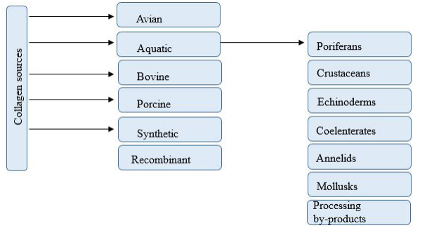 |
| Figure 1: Collagen sources |
Introduction
With an increased demand for polymers compatible with human tissue with no side and adverse effect on human body; biomaterials such as collagens have gained a great importance due to its potential to be used in different applications. Collagen forming 25% of the total body protein of vertebrates is a naturally occurring major connective tissue protein as well as bone, skin, cartilage, tendon and ligament [1. It compromises ¾ of the skin dry weight, and is the most abundant component in extracellular matrix [2, 3] (Table 1). So far twenty-eight different types of collagen composing 46 distinct polypeptide chains have been reported and identified in vertebrates [4].
Collagen has very critical role in the extracellular matrix especially in molecular and cellular interactions with the determination of both shape and form of the tissues. Importance of the collagen comes also from its important role in tissue and organ formation and cell expression. It is one of the best surface-active agent and has ability to penetrate a lipid-free interface. Compare to gelation and albumin known as natural biopolymers, collagen has higher biodegradability in biologic environment without leaving no permanent foreign residue, weak antigenity, and superior biocompatibility. Moreover, collagen has ability to form extra strong fiber and provide stability of the fiber by cross-linking and self-aggregation [5].
Collagen is one of the most suitable biomaterial in biomedical applications due to its safety and excellent biocompatibility, but it has poor mechanical strength and limited efficiency in infected sites [6. Thus, current use of collagen with future perspective need to be addressed in order to understand its function as biomaterial better.
Knowledge regarding biosynthesis, structure, and interactions of collagen would help development of different biomaterials to be used for burn treatment, bone repair, wound closure, hernia repair, hemostasis, cartilage defects, and various dental applications [7, 8]. Collagen use in drug delivery systems includes gel form combined with liposomes, nanoparticles for gene delivery, tablets and pellets form for protein delivery, control material for transdermal delivery in addition to forming artificial blood vessels, skin replacement, and bone substitutes [6].
Collagen structure
Collagen is a rod-shaped molecule with 3000 Å in length 15 Å in width with 300 kDa molecular weight [6]. Collagen structure involves three polypeptide chain wrapped around another like three-stranded rope with each chain having individual twisting in opposite direction to each other. High glycine and amino acid residues effect formation of helix structure in collagen molecule [6]. Collagen molecules can contain identical (homomeric) or genetically distinct (heteromeric) α chains. Each strand in collagen molecule is initially formed as a left-handed symmetry before its structure is finalized as a right-handed triple helix [9] (Figure 1).
The triple helix stability is provided by intra- and inter-molecular hydrogen bonds associated with glycyl residue of NH and CO groups in the neighboring chain. Bond formation also occurs by introduction of water molecules though the formation of hydrogen bonds with the help of the collagen hydroxyl groups [10]. Collagen family due to differences in the length of helix, presence of non-helical components in the structure, variations in the structure of the basic polypeptide chain, interruptions in helix structure, and differences in the termination of the helical domain has been classified into different groups [11]. These structural differences allow collagen structures to be classified as FACIT (Fibril Associated Collagens with Interrupted Triple Helices), FACIT-like collagen, fibrillar collagens, basement membrane collagen, beaded filament collagen, transmembrane collagen, short-chain collagen, and unclassified collagen. Different type of collagens differ in their length of the helix and fractions of non-helical components [12, 13].
Depending on their structure, more than 20 different types of collagen have been characterized with triple helix conformation having three polypeptide chain twined around each other. Repeated triplets in the each chain of the right-handed helical structure contain repeated sequence of glycine-X-Y with X and Y are indicated as proline or hydroxyproline, consequently [10]. Glycine is buried inside the core of the rod-shaped structure; whereas the other amino acids are placed on the surface [9]. This structure is further supported by interchain N-H (Gly) O=C(x) hydrogen bonds and electrostatic interactions [14]. Presence of triple helix is the main property of the collagen structure. Triple helix structure may vary depending on the type of the collagen present in the structure [11.
Type I collagen classified as fibrillar group synthesized as procollagen (soluble precursor molecules) is the most common form. Stability of fibrillary collagen is greatly depended on non-reducible covalent cross-linking in the triple helix [15]. Fibrils in FACIT collagen is interrupted by non-helical domains, and compare to fibril forming collagens C-NC domain in these molecules are shorter. Type IX, XII, XIV, XVI, XIX, XX, XXI, and XXI are also belong to FACIT group. The collagen molecules in the beaded filament collagen group constructed with no cleavage in the terminal region, and these uncleaved regions facilitate the bead region in collagen filaments. The most distinct characteristic of FACIT group is association of large N- and C-terminals [9, 12, 16] like in type VI even though its short triple helical domain. Moreover, type VI collagen is also classified under beaded filament collagen [9]. Non-fibrillar collagen includes the group of the basement membrane and associated collagen; whereas collagen IV is classified under non-fibrillar collagen present in epithelial lining cavities, endothelium tissue in blood vessel, muscle, nerve, and fat cells. Collagen IV which molecules are longer compare to fibrillar collagen is classified under non-fibrillar collagen subgroup that is present as thin sheets [9]. Collagen group VII is essential for functional integrity [11]. Short chain collagen locating under endothelial cells are defined as mesh forming collagen. Mineralizing cartilage contain some of the short chain collagens which also include type VIII functioning in cell proliferation as a growth enhancer and X group of collagens [12].
Transmembrane collagens having long but interrupted triple helical domain with a short N terminal domain act as cell surface receptors and matrix components involved in adhesion [9, 12, 17, 18]. Type XIII, XVII, XXIII, XXV, and the other collagen-like proteins are classified as transmembrane collagens. Type I, II, III, V, and XI are distinct with their three chains and continuous triple-helical structure. Except for type XI, the others are defined as fibril-forming collagens. Besides, they carry large sections of homologous sequences. The triple helical conformation regions in the type IV collagen are interrupted with large non-helical domains and with the short non-helical peptide interruption. Type IX, XI, XII, and XIV collagens are also known as fibril associated with small chains containing non-helical domains [17, 19]. Type VI and type VII collagens are classified as microfibrilla and fibril collagens, respectively. Triple helix topocollagen molecules form a fibril structure with distinct periodicity are the result of orderly arrangement. Both ends of the molecules have connected with nonhelical telopeptides that serve as the main source of antigenicity. After the discovery of type III collagen containing type I collagen and forming mixed fibrils as well as discovery of type IV, the terminology of the collagen was reorganized. Collagens were numbered with Roman numbers (I, II, III, IV, etc); whereas polypeptide chain are classified using α chains with Arabic numbers (α1, α2, α3, etc.) [17, 19] (Table 2).
Collagen sources
Slaughterhouse by products such as bones, hides, cartilages, tendons, or recombinant collagen are commonly used as raw material for collagen production with bovine and pigs as primary source at industrial scale [20]. Collagen utilized in cosmetics, medicine and other non-biomedical applications are usually use bovine collagen. Sterile and purified collagen from cow skin is usually utilized as injectable bovine collagen [21]. Both pig skin and bones are commonly used for porcine collagen extraction. Pig hides are also used for porcine type I collagen source [22, 23]. Poultry by-products such as bones, cartilage, and skin, although less preferred are also utilized as collagen source [24].
Annelids, poriferans, echinoderms, coelenterates, crustaceans, and mollusks have been widely utilized as collagen source [25, 26]. Japanese sea bass skin, yellow fine tuna bladder, skins, fin, bones and swim bladder of big head carp, sea urchin, cartilage from sturgeon [ 27] and sponges, and sea cucumber are the some of the marine originated collagen sources [26-30]. Type I collagen mainly extracted from Japanese sea-bass [31], sole fish [32], skin of silver carp [33], bullhead shark [34],mackerel [35], scales of Nile tilapia [28], and from the bones of skipjack tuna [36] (Fig.1).
Properties of collagen
Functional properties of collagen can be categorized as gelling behaviors of texturing, gel formation, water binding capacity, thickening and surface behaviors of foaming and stabilization, colloid function, emulsification, film formation, and colloid function [20, 37]. Collagen molecules can form aggregation with the cleavage of intra- and inter-molecular crosslinks during thermal solubilization. Gel forming ability of collagen can change depending on the type of collagen, ionic strength, and pH [38, 39].
Gel melting point and gel strength are the most important properties of the gels as they are extensively used in food, pharmaceutical, and cosmetic industries [40]. Collagen with its hydrophilic nature is used for the minimization of dripping loss on meat and fish products. Gelling properties of collagen, on the other hand, made it possible to be used as wetting agents in food, medical, and pharmaceutical applications [20, 40]. Water absorption property of collagen obtained from sea cucumber Stichopus japonicas improves the appearance, as well as the physical and sensory properties of the final products after thermal treatment [41. Presence of the polar in addition to carboxyl (-COOH) and hydroxyl (-OH) groups on the surface of the collagen molecule obtained from sea cucumber Holothuria cinerascens is important feature to improve the moisture retention of products especially in cosmetic products [42].
Charged groups of collagen having hydrophilic or hydrophobic amino acids determine its surface properties. Both hydrophilic or hydrophobic groups have the ability to reduce surface tension, and hydrophilic areas of the peptide chain have great impact on foaming and emulsifying properties [43].There is a positive correlation between the emulsion capacity of the collagen and protein concentration as well as hydrophobic amino acid content. Moreover, foaming and emulsifying properties of collagen is affected by pH, temperature, homogenization, and concentration [44, 45].
Common use of collagen
Production of film/disc/sheet for treatment of infections in different tissues such as liver cancer or infected corneal tissue as well as drug carrier for different antibiotics are common use of collagen. Collagen can form film with 0.01-0.5 mm thickness and drug can be loaded into the collagen membrane by either simple entrapment, covalent and/or hydrogen bonding. Collagen film are useful as gene delivery tool to promote bone formation. Collagen film and/or matrices can also act as scaffolds to help survival and growth of the transfected fibroblasts [46].
Collagen-based drug delivery systems are useful tools to heal corneal epithelium from the blinking action of the eyelids. Collagen matrix can serve as drug reservoir as drugs are entrapped inside the collagen matrix. Eye tear flushing though the shield provides dissolving the shield allowing to lubricate eye surface and increase healing of the epithelial [47].
Collagen sponges are combinedwith elastin, glycosaminoglycans or fibronectin to increase their fluid building capacity and elasticity. Moreover, crosslinking with glutaraldehyde, copolymerization with other polymers such as polyhydroxy ethyl methacrylate (PHEMA) ameliorate the collagen membrane properties of tensile strength as well as increase in the healing efficacy of infected wounds and burns. Collagen sponges are very effective to heal different type of dermal and epidermal wounds, absorb large amount of tissue exudates, and shield against secondary bacterial infections [48, 49].
Collagen gels are good drug delivery matrices and used as injectable collagen [50]. (Uchio et al., 2000). Gel produced with atelocollagen is used to repair cartilage defects by acting as carrier for chondrocytes. Combination of collagen with PHEMA as hydrogel has been formulated as delivery systems for anticancer drugs such as 5-FU [49].
Developed minipellet made from collagen is very small that can be injected through a syringe needle yet spacious enough to carry big molecular weight proteins. Different minipellets with different carrier compounds have been developed. Among them, collagen-based pellet designed as gene delivery tool is studied substantially [51].
Nanoparticles with sodium sulfate as dissolving agents provide charge-charge interactions between DNA and collagen. Moreover, nanoparticles are useful tools as a parenteral carrier for cytotoxic agents and therapeutic compounds like hydrocortisone and campthocin as well as anti HIV drugs [49].
Collagen, because of its ability to control evaporation of the fluid, keep wound flexible and pliable, develop granulation tissue, and provide mechanical protection against bacterial attack; is widely used for treatment of wounds. Because of its excellent adhesion to the wound and stimulation of cell reactivity [48], it is used in dental therapy. It prevents clot formation but epithelial cell rejuvenation, neovascularization, and stabilization of the connective tissue.
Films made from hydrolyzed collagen have been used as a tissue adhesive for future replacement due to its chemical resemblance to connective tissue and tissue fluid-binding properties. Moreover, it is biodegradable, non-toxic, and readily absorbed; therefore, it does not impose a hindrance to the healing process [52]. Cell culture mixed with homogenized reconstituted collagen is used for endodontic repair [52].
Collagen use in food and beverages
Since production of collagen decreases with age and bad diet, recent studies has focused on collagen addition to food products to gain collagen by diet as some people do not prefer to get injections. Food grade bovine (Colageno), type I and III ovine collagen (Ovinex), marine collagen (NiKollagen) are some examples of collagen supplementssuggested to be used as dietary supplements. Moisture absorption property of collagen as well as its fractions serve as nutritive fiber and protein source in human diets [53].
If collagen synthesis decreases, tissues are becoming thinner, weaker, and lose elasticity as human get older. Collagen supplements provide upholding the skin, nails, hair, and body tissues by attracting fibroblasts that synthesize new collagen. By developing collagen fibrils in the dermis and cohesion of the dermal collagen fibers, suppleness, thickness and resilience in addition to hydration of the tissues are improved and enhanced. Hydration of skin tissue had direct correlation with smoothness and reduction of wrinkling. Collagen use can increase muscle gain, reconstruct damaged joint, decrease time for recovery, and provide improvement in cardiovascular performance by virtue of creatine. Moreover, arginine present in collagen help to increase muscle mass [54]. Type II collagen is suggested to be used for rheumatoid arthritis treatment, swelling and stiffness of joints, and a chronic inflammatory sickness [55]. In addition, type II collagen use for 6 months helps to reduce morning stiffness, pain, tender joint, and swollen joint.
Collagen can also be used as food additive for the improvement of texture, color, flavor, and quality. The use of collagen in sausages and frankfurters provides improvement of rheological properties and nutritive value [53]. Heat treated collagen fiber can be used as emulsifier to increase protein solubility in water by decreasing oil-protein interactions. Heat treated collagen fiber provides higher creaming index and increase in the rate of emulsion. Similarly, addition of food grade collagen to bologna formulation [56], duck feet collagen addition to threadfin bream and sardine surimi, and chicken feet collagen addition to jelly production [57] are successful examples of collagen use in food formulations.
Use of biodegradable film as a replacement of plastics are getting more attention in food and cosmetic industry. Gelatin haslimited use as biodegradable film due to its hygroscopic nature; thus studies focus on the potential of collagen as biodegradable film [58]. Film forming properties of collagen and collagen-based derivatives are correlated with their amino acid composition and molecular weight distribution that are related to mechanical and barrier properties of the biodegradable film [59]. Collagenbased edible films and coating may be applied to different food by brushing, immersing, or spraying to protect foods against oxygen, moisture, and solute migration, as well as to provide structural integrity [56]. Collagen films or coatings are able to carry active substances of colors, antioxidants, flavors and antimicrobials [60, 61] as well as rosemary extract in processed meat preparation [62].
Beside food formulations, collagen addition is practiced to sausage casing [53], netted roasts, fish fillets, boneless hams, meat pastes, and roast beef [56]. Soy collage, cappuccino collagen, and cocoa collagen are some examples of the collagen addition to drinks and collagen-infused drinks as well as energy drinks. It is reported that collagen drink stimulates the body collagen mechanism resulting in sagging and skin wrinkles. Production of marine fish peptide collagen drink from salmon fish skin [56], coffee formulations with collagen from fish source [63], and bovine collagen in refining beer and yeast [64] are the successful formulations.
Conclusions
Collagen -with different superior properties- finds its place in variety of industries as biomaterial or ingredient in formulations. It can be used solely and/or combination with different compounds in different biomaterials and products. Studies mostly focused either on health promoting effect of collagen like medical and pharmaceutical uses or improvement of functional and quality properties like uses in food and drink formulations. However, studies regarding stability of collagen in different use, their degradation kinetics and factor affecting stability of collagen in different formulation still need to be searched in future studies.
Conflict of interest
The authors declare no conflict of interest.
References
- Ramshaw, J.A.M. (2016). Biomedical applications of collagens. Journal of Biomedical Materials Research Part B: Applied Biomaterials, 104, 665–675.
- Brinckmann, J. (2005). Collagens at a Glance. In: Collagen: Primer in Structure, Processing and Assembly, Topics in Current Chemistry (edited by J. Brinckmann, H. Notbohm & P.K. Müller). Pp. 1–6. Berlin, Heidelberg: Springer.
- Shoulders, M.D. & Raines, R.T. (2009). Collagen structure and stability. Annual Review of Biochemistry, 78, 929–958.
- Veit, G., Kobbe, B., Keene, D.R., Paulsson, M., Koch, M. & Wagener, R. (2006). Collagen XXVIII, a Novel von Willebrand Factor a domain-containing protein with many imperfections in the collagenous domain. Journal of Biological Chemistry, 281, 3494–3504.
- Maeda, M., Tani, S., Sano, A. & Fujioka, K. (1999). Microstructure and release characteristics of the minipellet, a collagen-based drug delivery system for controlled release of protein drugs. Journal of Controlled Release, 62, 313–324.
- Lee, C.H., Singla, A. & Lee, Y. (2001). Biomedical applications of collagen. International Journal of Pharmaceutics, 221, 1–22.
- Antoine, E.E., Vlachos, P.P. & Rylander, M.N. (2014). Review of collagen I hydrogels for bioengineered tissue microenvironments: characterization of mechanics, structure, and transport. Tissue Engineering Part B: Reviews, 20, 683–696.
- Miranda-Nieves, D. & Chaikof, E.L. (2017). Collagen and elastin biomaterials for the fabrication of engineered living tissues. ACS Biomaterials Science & Engineering, 3, 694–711.
- Feyzi, S., Varidi, M., Zare, F. & Varidi, M.J. (2015). Fenugreek (Trigonella foenum graecum) seed protein isolate: extraction optimization, amino acid composition, thermo and functional properties. Journal of the Science of Food and Agriculture, 95, 3165–3176.
- Habermehl, J., Skopinska, J., Boccafoschi, F., Sionkowska, A., Kaczmarek, H., Laroche, G. & Mantovani, D. (2005). Preparation of ready-to-use, stockable and reconstituted collagen. Macromolecular Bioscience, 5, 821–828.
- Wang, L., Liang, Q., Chen, T., Wang, Z., Xu, J. & Ma, H. (2014). Characterization of collagen from the skin of Amur sturgeon (Acipenser schrenckii). Food Hydrocolloids, 38, 104–109.
- Sherman, V.R., Yang, W. & Meyers, M.A. (2015). The materials science of collagen. Journal of the Mechanical Behavior of Biomedical Materials, SI:Collagen mechanics, 52, 22–50.
- Terzi, A., Gallo, N., Bettini, S., Sibillano, T., Altamura, D., Campa, L., Natali, M.L., Salvatore, L., Madaghiele, M., De Caro, L., Valli, L., Sannino, A. & Giannini, C. (2019). Investigations of processing–induced structural changes in horse type-I collagen at sub and supramolecular levels. Frontiers in Bioengineering and Biotechnology, 7, 203.
- Persikov, A.V., Ramshaw, J.A.M. & Brodsky, B. (2005). Prediction of collagen stability from amino acid sequence. Journal of Biological Chemistry, 280, 19343–19349.
- Kadler, K.E., Baldock, C., Bella, J. & Boot-Handford, R.P. (2007). Collagens at a glance. Journal of Cell Science, 120, 1955– 1958.
- Mizuno, K., Boudko, S.P., Engel, J. & Bächinger, H.P. (2010). Kinetic hysteresis in collagen folding. Biophysical Journal, 98, 3004–3014.
- Ertem, F., Watson, A., Ramos- Rivers, C., Babichenko, D., Tang, G., Schwartz, M., Proksell, S., Johnston, E., Hashash, J., Barrie, A., Harrison, J., Koutroubakis, I., Dunn, M., Hartman, D. & Binion, D. (2019). Mo1823 – Endoscopic patterns and location of post-operative recurrence in crohn’s disease patients with side to side anastomosis following ileocecal resection. Gastroenterology, 156, 850.
- Senadheera, T.R.L., Dave, D. & Shahidi, F. (2020). Sea cucumber derived type I collagen: A comprehensive review. Marine Drugs, 18, 471.
- Franzke, C.-W., Bruckner, P. & Bruckner-Tuderman, L. (2005). Collagenous transmembrane proteins: Recent insights into biology and pathology. The Journal of Biological Chemistry, 280, 4005–8.
- Gómez-Guillén, M.C., Giménez, B., López-Caballero, M.E. & Montero, M.P. (2011). Functional and bioactive properties of collagen and gelatin from alternative sources: A review. Food Hydrocolloids, 25 years of Advances in Food Hydrocolloid Research, 25, 1813–1827.
- Rizk, M.A. & Mostafa, N.Y. (2016). Extraction and characterization of collagen from buffalo skin for biomedical applications. Oriental Journal of Chemistry, 32, 1601–1609.
- Chen, X., Wu, J., Li, L. & Wang, S. (2017). The cryoprotective effects of antifreeze peptides from pigskin collagen on texture properties and water mobility of frozen dough subjected to freeze–thaw cycles. European Food Research and Technology, 243, 1149–1156.
- Maione-Silva, L., Castro, E.G. de, Nascimento, T.L., Cintra, E.R., Moreira, L.C., Cintra, B.A.S., Valadares, M.C. & Lima, E.M. (2019). Ascorbic acid encapsulated into negatively charged liposomes exhibits increased skin permeation, retention and enhances collagen synthesis by fibroblasts. Scientific Reports, 9, 522.
- Munasinghe, K.A., Schwarz, J.G. & Whittiker, M. (2015). Utilization of Chicken By-Products to Form Collagen Films. Journal of Food Processing, 2015, e247013.
- Dave, D., Liu, Y., Clark, L., Dave, N., Trenholm, S. & Westcott, J. (2019). Availability of marine collagen from Newfoundland fisheries and aquaculture waste resources. Bioresource Technology Reports, 7, 100271.
- Shahidi, F., Varatharajan, V., Peng, H. & Senadheera, R. (2019). Utilization of marine by-products for the recovery of value-added products. Journal of Food Bioactives, 6.
- Kaewdang, O., Benjakul, S., Kaewmanee, T. & Kishimura, H. (2014). Characteristics of collagens from the swim bladders of yellowfin tuna (Thunnus albacares). Food Chemistry, 155, 264– 270.
- Kittiphattanabawon, P., Benjakul, S., Sinthusamran, S. & Kishimura, H. (2015). Characteristics of collagen from the skin of clown featherback (Chitala ornata). International Journal of Food Science & Technology, 50.
- Manchinasetty, N.V.L., Oshima, S. & Kikuchi, M. (2017). Preparation of flexible bone tissue scaffold utilizing sea urchin test and collagen. Journal of Materials Science: Materials in Medicine, 28, 184.
- Song, H. & Li, B. (2017). Beneficial effects of collagen hydrolysate: A review on recent developments. Biomedical Journal of Science and Technology Research, 1, 1–4.
- Kim, H.K., Kim, Y.H., Kim, Y.J., Park, H.J. & Lee, N.H. (2012). Effects of ultrasonic treatment on collagen extraction from skins of the sea bass Lateolabrax japonicus. Fisheries Science, 78, 485– 490.
- Arumugam, G.K.S., Sharma, D., Balakrishnan, R.M. & Ettiyappan, J.B.P. (2018). Extraction, optimization and characterization of collagen from sole fish skin. Sustainable Chemistry and Pharmacy, 9, 19–26.
- Abdollahi, M., Rezaei, M., Jafarpour, A. & Undeland, I. (2018). Sequential extraction of gel-forming proteins, collagen and collagen hydrolysate from gutted silver carp (Hypophthalmichthys molitrix), a biorefinery approach. Food Chemistry, 242, 568–578.
- Berillis, P. (2015). Marine collagen: Extraction and applications. Research Trends in Biochemistry, Molecular Biology and Microbiology, 1-13.
- Hashimoto, K., Kobayashi, S. & Yamashita, M. (2017). Comparison of connective tissue structure and muscle toughness of spotted mackerel Scomber australasicus and Pacific mackerel S. japonicus during chilled and frozen storage. Fisheries Science, 83, 133–139.
- Yu, D., Chi, C.-F., Wang, B., Ding, G.-F. & Li, Z.-R. (2014). Characterization of acid-and pepsin-soluble collagens from spines and skulls of skipjack tuna (Katsuwonus pelamis). Chinese Journal of Natural Medicines, 12, 712–720.
- Coppola, D., Oliviero, M., Vitale, G.A., Lauritano, C., D’Ambra, I., Iannace, S. & Pascale, D. de. (2020). Marine collagen from alternative and sustainable sources: extraction, processing and applications. Marine Drugs, 18, E214.
- Sadowska, M., Kołodziejska, I. & Niecikowska, C. (2003). Isolation of collagen from the skins of Baltic cod (Gadus morhua). Food Chemistry, 81, 257–262.
- Liu, Z., Oliveira, A.C.M. & Su, Y.-C. (2010). Purification and characterization of pepsin-solubilized collagen from skin and connective tissue of giant red sea cucumber (Parastichopus californicus). Journal of Agricultural and Food Chemistry, 58, 1270–1274.
- Karim, A.A. & Bhat, R. (2009). Fish gelatin: properties, challenges, and prospects as an alternative to mammalian gelatins. Food Hydrocolloids, 23, 563–576.
- Dong, X., Zhu, B., Sun, L., Zheng, J., Jiang, D., Zhou, D., Wu, H. & Murata, Y. (2011). Changes of collagen in sea cucumber (Stichopus japonicas) during cooking. Food Science and Biotechnology, 20, 1137.
- Li, P.-H., Lu, W.-C., Chan, Y.-J., Ko, W.-C., Jung, C.-C., Le Huynh, D.T. & Ji, Y.-X. (2020). Extraction and characterization of collagen from sea cucumber (Holothuria cinerascens) and its potential application in moisturizing cosmetics. Aquaculture, 515, 734590.
- Du, Y.-N., Guo, X.-K., Han, Y.-T., Han, J.-R., Yan, J.-N., Shang, W.-H. & Wu, H.-T. (2019). Physicochemical and functional properties of protein isolate from sea cucumber (Stichopus japonicus) guts. Journal of Food Processing and Preservation, 43, e13957.
- Abdelmalek, B.E., Gómez-Estaca, J., Sila, A., Martinez-Alvarez, O., Gómez-Guillén, M.C., Chaabouni-Ellouz, S., Ayadi, M.A. & Bougatef, A. (2016). Characteristics and functional properties of gelatin extracted from squid (Loligo vulgaris) skin. LWT - Food Science and Technology, 65, 924–931.
- Bhuimbar, M.V., Bhagwat, P.K. & Dandge, P.B. (2019). Extraction and characterization of acid soluble collagen from fish waste: Development of collagen-chitosan blend as food packaging film. Journal of Environmental Chemical Engineering, 7, 102983.
- Park, J.C., Hwang, Y.S., Lee, J.E., Park, K.D., Matsumura, K., Hyon, S.H. & Suh, H. (2000). Type I atelocollagen grafting onto ozone-treated polyurethane films: Cell attachment, proliferation, and collagen synthesis. Journal of Biomedical Materials Research, 52, 669–677.
- Leaders, F.E., Hecht, G., VanHoose, M. & Kellog, M. (1973). New polymers in drug delivery. Annals of Ophthalmology, 5, 513–516.
- Chvapil, M., Kronenthal, R.L. & Winkle, W. van. (1973).Medical and Surgical Applications of Collagen. In: International Review of Connective Tissue Research (edited by D.A. Hall & D.S. Jackson). pp. 1–61. Elsevier.
- Khan, R. & Khan, Mohd.H. (2013). Use of collagen as a biomaterial: An update. Journal of Indian Society of Periodontology, 17, 539–42.
- Uchio, Y., Ochi, M., Matsusaki, M., Kurioka, H. & Katsube, K. (2000). Human chondrocyte proliferation and matrix synthesis cultured in Atelocollagen® gel. Journal of Biomedical Materials Research, 50, 138–143.
- Nakagawa, T. & Tagawa, T. (2000). Ultrastructural study of direct bone formation induced by BMPs-collagen complex implanted into an ectopic site. Oral Diseases, 6, 172–179.
- Bashutski, J.D. & Wang, H.-L. (2009). Periodontal and endodontic regeneration. Journal of Endodontics, 35, 321–328.
- Neklyudov, A.D. (2003). Nutritive fibers of animal origin: Collagen and its fractions as essential components of new and useful food products. Applied Biochemistry and Microbiology, 39, 229–238.
- King’ori, A. (2011). A Review of the uses of poultry eggshells and shell membranes. International Journal of Poultry Science, 10, 908–912.
- Zhang, L., Xu, W., Yue, P., Wang, Q., Li, Y., Pei, X. & Zeng, P. (2020). High occurrence of aflatoxin B1 in Pixian Doubanjiang, a typical condiment in Chinese cuisine. Food Control, 110, 107034.
- Hashim, P., Mohd Ridzwan, M.S., Bakar, J. & Mat Hashim, D. (2015). Collagen in food and beverage industries. International Food Research Journal, 22, 1–8.
- Almeida, P.F. de, Araújo, M.G.O. de & Santana, J.C.C. (2012). Collagen extraction from chicken feet for jelly production. Acta Scientiarum Technology, 34, 345–351.
- Ben Slimane, E. & Sadok, S. (2018). Collagen from cartilaginous fish by-products for a potential application in bioactive film composite. Marine Drugs, 16, 211.
- Teixeira, B., Marques, A., Pires, C., Ramos, C., Batista, I., Saraiva, J. & Nunes, M. (2014). Characterization of fish protein films incorporated with essential oils of clove, garlic and origanum: Physical, antioxidant and antibacterial properties. LWT - Food Science and Technology, 59(1), 533-539.
- Bourtoom, T. (2008). Edible films and coatings: Characteristics and properties. International Food Research Journal, 15(3), 237-248.
- Suput, D., Lazic, V., Popovic, S. & Hromis, N. (2015). Edible films and coatings: Sources, properties and application. Food and Feed Research, 42, 11–22.
- Waszkowiak, K. & Dolata, W. (2007). The application of collagen preparations as carriers of rosemary extract in the production of processed meat. Meat Science, 75, 178–183.
- Yacoubou, J. (2011). Nestle Malaysia collagen containing Nescafe Body Partner coffee discontinued: Update. Downloaded from http:// www.vrg.org/blog/2011/12/15/nestle-malaysiacollagen-containing-nescafe-body-partner-coffeediscontinued-update/on 5/9/2012.
- Hickman, D., Sims, T.J., Miles, C.A., Bailey, A.J., Mari, M. de & Koopmans, M. (2000). Isinglass/collagen: denaturation and functionality. Journal of Biotechnology, 79, 245.
Artcle Information
Review Article
Received Date: May 01, 2025
Accepted Date: May 17, 2025
Published Date: May 24, 2025
Journal of Foodscience Nutrition and Public Health
Volume 1 | Issue 1
Citation
Gulsun Akdemir Evrendilek (2025) Collagen: Structure, Synthesis and Common Use. J Food Sci Nutr Public Health 1: 105
Copyright
©2025 Gulsun Akdemir Evrendilek. This is an open-access article distributed under the terms of the Creative Commons Attribution License, which permits unrestricted use, distribution, and reproduction in any medium, provided the original author and source are credited.

doi: jfnp.2025.1.105