

Review Article
Volume-1 Issue-1, 2025
Surgical Management of Colovesical Fistula at A Public Hospital In Southeastern Mexico
-
Received Date: May 05, 2025
-
Accepted Date: May 24, 2025
-
Published Date: May 31, 2025
Journal Information
Abstract
Introduction: Diverticular disease of the colon preferentially affects older adults, with 10%-25% developing complications that include colovesical fistula. The fact that there are few reports on its management in Mexico motivated us to carry out the present study.
Aim: To evaluate the results of surgical treatment of colovesical fistulas at a hospital in Southeastern Mexico.
Material and Methods: A retrospective and comparative study was conducted on patients seen at an advanced specialty hospital in Veracruz, that were divided into 2 groups: A) conventional surgery 86.36% and B) laparoscopic surgery 13.64%.
Variables Analyzed : Age, sex, risk factors, type of surgery, surgery duration, hospital stay, complications, and mortality
Statistical analysis: The quantitative variables were calculated through descriptive statistics and the continuous variables through the Student’s t test, utilizing IBM-SPSS, version 25.0, software for Windows.
Results: The cohort consisted of 44 patients with a mean age of 62.12 ± 12.25 years, a predominance of women 56.82%, and a mean body mass index of 31.82±5.89 kg/m2. Of the patient total, 88.64% had comorbidities and the anesthesia risk grade was I-II. The differences regarding anesthesia/surgery duration and intraoperative bleeding were statistically significant between the two groups, whereas the differences in days of hospital stay, complications, and reinterventions were not. Oral diet resumption, length of follow-up, and bladder catheter removal were similar between the groups and there were no deaths.
Discussion: Our results were satisfactory and comparable to those published by other authors. The two approaches are equally safe, and the laparoscopic approach has advantages over open surgery.
Key words
Complicated Diverticular Disease; Colovesical Fistula; Conventional Approach; Laparoscopic Approach
Parameter |
n = 44 |
% |
Sex |
25 |
56.82 |
Mean age (years) |
62.12 ± 12.21 |
(34-98) |
Mean body mass index (kg/m2) |
31.82 ± 5.89 |
(18-38) |
Progression time (weeks) |
15.02 ± 5.02 |
(8-48) |
Associated comorbidities |
39 |
88.64 |
General risk (ASA) |
16 |
36.36 |
Parameter |
Group A |
Group B |
p |
Anesthesia/surgery duration (min) |
220.08 ± 44.7 |
340.23 ± |
0.001 |
Intraoperative bleeding (ml) |
332.45 ± 14.3 |
168.1 ± 12.8 |
0.001 |
Conversion rate |
- |
0 (0.00) |
- |
Mean days of hospital stay |
9.92 ± 19.7 |
4.42 ± 0.5 |
0.228 |
Prophylactic ileostomy |
3 (7.89) |
0 |
0.183 |
Oral diet resumption (hours) |
48.71 ± 8.9 |
45.62 ± 4.7 |
0.165 |
Bladder catheter removal (weeks) |
32.63 ± 5.8 |
31.4±3.5 |
0.037 |
Length of follow-up (months) |
14.94 ±13.6 |
13.82 ±4.3 |
0.235 |
Complications |
10 (26.32) |
2 (33.33) |
0.082 |
Reintervention |
5 (13.16) |
- |
0.068 |
Mortality |
0 |
0 |
- |
Author |
Total/ Open/ |
Preventive ileostomy |
Conversion rate |
Clavien-Dindo III-IV |
Reintervention |
n /(%) |
n / (%) |
(%) |
n / (%) |
n / (%) |
|
Bartus C. (15) |
40 (100.00) |
5(12.5%) |
1 (25.00) |
2 (5.00%) |
0 (0.00) |
Garcea G. (21) |
64 (100.00) |
11(17.18) |
3(15.78) |
1 (1.67) |
0 (00.00) |
Marney L A. (24) |
15 (100.00) |
0 (0.00) |
5 (33.33%) |
1(6.67) |
0 (0.0) |
Maciel V. (25) |
55 (100.00) |
5 (9.09) |
8 (14.55) |
5 (9.09) |
0 (0.00) |
Badic B. (26) |
37 (100.00) |
5 (13.51) |
3 (17.65) |
3 (8.11) |
0 (0.00) |
Marcucci T. (12) |
16 (100.00) |
1 (6.25) |
2(12.50) |
5 (31.25) * |
2(12.50) |
De León M. (9) |
52 (100.00) |
6 (11.54) |
6 (20.69) |
5 (9.61) |
0 (0.00) |
Martinolich J. |
52 (100.00) |
5 (9.61) |
18 (34.61) |
14 (26.92) * |
0 (0.00) |
De la Fuente H. |
13 (100.00) |
0 (0.00) |
3 (33.33) |
1(11.1) |
0 (0.00) |
Kitaguchi D.(28) |
11(100.00) |
0 (0.00) |
3 (27.00) |
1(0.09) |
0 (0.00) |
Weng Y (29) |
7 (100.00) |
0 (0.00) |
2 (40.00) |
1(14.28) |
0 (0.00) |
Tomizawa K (30) |
39 (100.00) |
0 (0.00) |
0 (0.00) |
0 (0.00) |
0 (0.00) |
Total |
401 (100.00) |
38 (9.47) |
54(21.95) |
39 (7.23) |
2 (0.50) |
Carrasco A. M. |
44 (100.00) |
3 (8.82) |
(0.00) |
0 (0.00) |
2 (4.54) |
*Not classified.
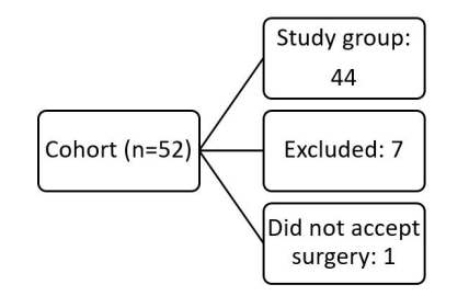 |
| Graph 1: Application of the selection criteria of the cohort of cases |
Introduction
Colovesical fistula (CVF) is the pathologic communication between the sigmoid colon and the urinary bladder, the most common cause of which tends to be diverticular disease, accounting for 50-88% of cases. However, it can also present in other diseases, such as colon cancer (10-12%), inflammatory bowel disease (5-8%), non-Hodgkin lymphoma and AIDS, as well as resulting from radiotherapy sequelae, trauma lesions, and iatrogenic lesions during pelvic surgery (4-13%), with resolution requiring surgical management [1].
The frequency of CVF in complicated diverticular disease has been estimated at 2-6%, with a range of 2-23%. That is especially true in older adult patients, in whom risk factors for chronic-degenerative diseases tend to condition morbidity in 16-35% of patients [1-5] with a worldwide mortality rate of 0.51±0.31/100.000 and a range of 0.11-1.75 [6-8].
Surgical treatment consists of resecting the affected sigmoid colon, with primary anastomosis and closure of the lesion in the bladder wall, and should be performed as an elective procedure [9-11]. The anastomosis should be protected through ileostomy, if the surgeon feels there is the possibility of anastomotic dehiscence due to suture tension or technical difficulties during the surgery [12,13]. Conventional and laparoscopic are the chosen options, and Robotic assistance has recently been successfully introduced, but given its lack of availability and high cost, is only performed at national referral centers [14-21].
The results of surgical treatment are satisfactory in the majority of patients, but 2-23% tend to present with Clavien-Dindo III-V complications. Anastomotic dehiscence is the main complication, making reintervention necessary, and the mortality rate reported in the medical literature varies from 0 to 4,5%, especially when the disease is associated with advanced age and general risk factors [22].
In Mexico in 2007, it was published a 5-year experience regarding the management of cases treated at the Hospital General de México “Eduardo Liceaga” that underwent sigmoidectomy, with good results and no deaths and in Latin America, there are no reports on the results of surgical management of CVF, which is why we consider the present study to be important [23].
Materials and Method
A retrospective, descriptive, comparative study on a cohort of cases was conducted.
Study universe: Patients diagnosed with diverticular disease, complicated by colovesical fistula (CVF), treated at the UMAE No. 14 of the Centro Médico Nacional “Adolfo Ruiz Cortines” of the Instituto Mexicano del Seguro Social in Veracruz, within the time frame of 2017-2021.
Inclusion criteria: Adult subjects with CVF.
Exclusion criteria: Adult subjects with CVF whose medical records were incomplete.
Elimination criteria: Patients that did not accept surgical treatment.
Protocol design: Patients were clinically evaluated and routine laboratory studies, colonoscopy, double contrast CT and cardiopulmonary assessment were performed. All of them underwent mechanical cleansing through liquid diet and osmotic laxatives (Macrogol 105.00 gr. in 4 takes), the day before the intervention. Sigmoidectomy was performed, with primary anastomosis and bladder wall closure. In 3 cases (6.82%), defunctionalization was carried out through ileostomy, and hysterectomy was performed in the same surgical act in 8 cases (18.18%). The conventional and laparoscopic surgical approaches were performed. In the two approaches, two staplers were employed: a 60 mm linear stapler for the proximal closure of the rectal stump and a 29 or 33 mm stapler for the anastomosis. Bladder wall repair was carried out with continuous suturing, using synthetic absorbable 0 polyglycolic acid sutures, and finally underwent testing for anastomotic leakage, by insufflating pressurized air and compressing the proximal segment to verify its integrity. A simple Penrose drain, as well as a bladder catheter, were placed in 100% of the cases. The patients received a postoperative antimicrobial regimen, consisting of cefotaxime (n = 20; 45.45%), ciprofloxacin (n = 13; 29.55%), or ceftriaxone (n = 11; 25.00%), together with metronidazole, in all cases.
Variables analyzedAge, sex, progression time of the condition, risk factors, surgical approach, type of surgery performed, days of hospital stay, complications, and mortality.
Statistical analysisThe results were analyzed, using the descriptive statistics of measures of central tendency and dispersion for the quantitative variables and the Student’s t test for the continuous variables. The IBM-SPSS, version 25.0, software for Windows, was employed for the analysis.
Ethical aspectsThe present study was conducted according to the principles stated in the Declaration of Helsinki, the NOM-012-SSA3-2012, and the General Health Law for Health Research, article 17, which stipulates that the execution of research projects on humans must result in no risk for the study participants and that the data collected be completely confidential and anonymous. The protocol was approved by the Bioethics and Research Committee of the UMAE No 14 of the IMSS and the School of Medicine of the Universidad Veracruzana, Veracruz-Boca del Río.
Financial disclosureThe study was performed with resources from the participating institutions. There was no external funding.
Conflict of interestAll authors declare that there are no conflicts of interest.
Results
A total of 52 patients were studied. Seven of them were excluded due to incomplete medical records and 1 was eliminated for not accepting the surgical procedure, leaving a study group total of 44, graph 1.
The anthropometric characteristics of the entire study group were: mean age of 62,}.12±12,25 years (range 34-98), a predominance of women (n = 25; 56.82%) over men (n = 19; 43.18%), and a mean body mass index (BMI) of 31.82±5,89 kg/ m2 (range 18-38).
Progression time from disease onset to the performance of the intervention was 15.00±5.02 weeks (range 8-48). Thirty-nine cases (88.64%) presented associated comorbidities, and the ASA general anesthesia/surgery risks were: grade I and II in most of cases, table 1.
The patients were divided into two groups: group A) patients that underwent the conventional approach with open surgery (n = 38; 86.36%) and group B) patients that underwent the laparoscopic approach (n = 6; 13.64%).
Anesthesia/surgery durationIn group A, duration was 220.08±44.7 minutes (range 140-310), whereas in group B, it was 340,.23±14.3 minutes (range 140-310) (p=0,001).
Intraoperative bleedingIn group A, blood loss was estimated at 332.45±14.3 ml (rango100-500), and 1 patient required 2 units of packed red blood cells, whereas in group B, blood loss was 168.1±12.8 ml (range100-350) (p=0.006).
Conversion rateThere was no need for conversion to open surgery
DefunctionalizationIn group A, the surgeon decided on prophylactic defunctionalization in 3 cases (7.89%), whereas in group B it was not performed on any of the cases (p=0.183).
Oral diet resumptionIn group A, oral diet was resumed at 48.71±8.9 postoperative hours (range 24-72), whereas in group B, it was resumed at 45.62±4,7 postoperative hours (range 24-72) (p=0.235).
Hospital stayIn group A, the mean duration of hospital stay was 9.92±19,7 days (range 4-6), whereas in group B, it was 4.42±0,5 days (range 7-81) (p=0.228).
Bladder catheter removalIn group A, the catheter was removed on postoperative day 32.63±5,8, whereas in group B, removal was on postoperative day 31.4±3.5 (p=0.037).
Progression Length of follow-upIn group A, follow-up lasted 14.94±13.6 months, and in group B, it lasted 13.82±4,3 months (p=0.235).
ComplicationsIn group A, 10 cases (26.32%) presented with complications. In 5 of those cases (11.66%), they were mild Clavien-Dindo grade I complications, which included surgical wound infection (n = 4; 10.52%), seroma (n = 3; 7.89%), and anastomotic stricture (n = 1; 2.63%), all of which were resolved through conservative management. Two cases (5.26%) presented with Clavien-Dindo grade III complications, that included anastomotic leak and wall eventration, and required reintervention in the immediate postoperative period. In group B, there were only 2 ClavienDindo grade I complications: one case of seroma and one case of stricture (16.67%, respectively), which were resolved through conservative management. There was no grade III or grade IV complications (p=0.082) [1].
ReinterventionIn Group A, 5 cases (13.16%) underwent reintervention, 2 of which (5.26%) were performed in the immediate postoperative period. One of the cases (2.63%) of reintervention was due to anastomotic dehiscence, and ileostomy was carried out, and the other case was due to abdominal wall eventration. In the remaining 3 cases (7.89%) elective reintervention was performed six weeks after ileostomy closure. In group B, there were no reinterventions (p=0.068), table 2.
MortalityThere were no perioperative or postoperative deaths in either of the two groups.
Discussion
The standardized treatment of FVC is surgical resection of the sigmoid and suture of the vesical wall, which should be performed electively after the acute episode has been controlled. The open surgery approach can be carried out, although traditional, handassisted, or robotic-assisted laparoscopy can be performed at high-volume referral centers by experienced coloproctology surgeons. The laparoscopic techniques have shown results equal to those of the conventional approach, with less intraoperative blood loss, and significantly reduced morbidity, surgical site infections, and length of hospital stay. However, the use of laparoscopy has been the subject of debate due to the elevated conversion rate caused by the firm pelvic adhesions resulting from the inflammatory process [12-21].
The results of our cohort of cases were compared with eleven studies published over the past fifteen years, in which 401 patients that underwent surgical interventions (155 open surgeries [38.65%] and 246 laparoscopies [61,35%]) were analyzed [15, 21-30], table 4.
In the majority of the case series evaluated, the mean age was 68 years (range 28-84), whereas it was slightly lower, at 62.12±12.21 years (range 34-98), in our study. In general, male sex predominated (54-63%), whereas female sex predominated in our analysis (56.82%), with a BMI of 31.82±5.89 (range 18- 38). The comorbidities associated with complicated diverticular disease were frequent in all the case series. In our study, they presented in 88,26% of the patients, with the majority having one or two complications. The most frequent comorbidities were obesity, diabetes mellitus, and high blood pressure and the ASA risk was grade I and II in 90.91% of our patients, similar to that reported by different authors [26- 32].
Surgery duration in our study was a mean 220.08±44.7 minutes for open surgery, whereas it was 340.23± 14.3 minutes for the laparoscopic approach. The difference was statistically significant (p=0.001), similar to that published with a mean of 150 to 350 minutes for complicated diverticular disease, with longer times in cases of colovesical fistula [29-33].
Intraoperative blood loss was higher during open surgery, at 332.45±14.33 ml (range 100-500), with only one case requiring blood transfusion, whereas for the laparoscopic procedure, blood loss was only 168.1±12 ml, resulting in a statistically significant difference (p=0.001), higher than reported by other aurors of 75-125 ml [24,25,30] The use of preventive ileostomy is usually left up to the criterion and experience of the surgeon and is recommended in cases in which there is a possibility of anastomotic dehiscence, whether due to deficient tissue irrigation or suture line tension, we prefer the performance of the ileostomy as a defunctionalization procedure because its closure is simpler and with less morbidity than the transverse colostomy The results published by different authors is 9.47%, especially regarding open surgery, and is similar to the 7.89% of our open surgery cases (p=0.183) [9,12,14,15,21,24- 30]. The conversion rate to open surgery found in the literature is 21.95%, ranging from 0 to 40%, but in our case series, there was no need for conversion in any of the patients.
Immediate postoperative management was satisfactory in the majority of our patients. They all received an antimicrobial regimen made up of the combination of metronidazole with cefotaxime in 45.45%, ciprofloxacin in 29.55%, or ceftriaxone in 25,00%. There were no statistically significant differences, with respect to oral diet resumption, which took place at 48.71±8.9 hours after the open surgery and at 45.62±4.7 hours after the laparoscopic surgery (p=0.165), to the number of days of hospital stay, which was 9.92±19.7 days in the open surgery patients and 4.42±0.5 days in the laparoscopy patients (p=0.228), or to bladder catheter removal, which was on postoperative day 32.6±5.8 for open surgery and day 31.4±3.5 for the laparoscopic procedure (p=0.037).
The length of postoperative follow-up was similar in groups A and B (14.94±136 days and 13.82±4.3 days, respectively) (p=0.235). Complications presented in 10 cases (26.32%) in the open surgery group, 2 (5.26%) of which were Clavien-Dindo III (anastomotic leak and abdominal wall eventration) that required reintervention in the immediate postoperative period. In the laparoscopy group there were 2 minor complications (seroma and stricture) and no serious ones (p=0.082), similar to that reported in the universal literature (7.23%; range 0-23). The mortality rate was low in all the case series (0-2.3%) and there were no deaths in our study patients [9,12,14,15,21,24-30].
Conclusions
Sigmoidectomy with closure of bladder perforation is the recommended treatment in patients with colovesical fistula, which should be performed electively.
In our series as published by various authors we obtained good results with low morbidity and no mortality, so we consider it safe and reliable
Laparoscopic surgery has advantages over the conventional approach and is recommended to be performed in high-volume hospitals and by experienced surgeons.
References
- Biffoni M, Urciuoli P, Grimaldi G, Eberspacher C, Santoro A et al. (2019) Colovesical fistula complicating diverticular disease: diagnosis and surgical management in elderly. Minerva Chir 74: 187-88.
- Sell NM, Perez NP, Stafford CE, Chang D, Bordeianou LG et al. (2020) Are There Variations in Mortality From Diverticular Disease By Sex? Dis Colon Rectum 63: 1285-92.
- Kupcinskas J, Strate LL, Bassotti G, Torti G, Herszènyi L et al. (2019) Pathogenesis of Diverticulosis and Diverticular Disease. J Gastrointestin Liver Dis 28: 7-10.
- You H, Sweeny A, Cooper ML, Von Papen M, Innes J (2019) The management of diverticulitis: a review of the guidelines. Med J 211: 421-7.
- Rezapour M, Stollman N (2019) Diverticular Disease in the Elderly. Curr Gastroenterol Rep 21: 46.
- Hunt CW, Chaturvedi R, Brown L, Stafford C, Cauley CE et al. (2021) Diverticular Disease Epidemiology: Rising Rates of Diverticular Disease Mortality Across Developing Nations. Dis Colon Rectum 64: 81-90.
- Raña-Garibay R, Salgado-Nesme N, Carmona-Sánchez R, Remes-Troche JM, Aguilera-Carrera J et al. (2019) Consenso mexicano de diagnóstico y tratamiento de la enfermedad diverticular del colon. Rev Gastroenterol Mex 84: 220-40.
- Biffoni M, Urciuoli P, Grimaldi G, Eberspacher C, Santoro A (2019) Colovesical fistula complicating diverticular disease: diagnosis and surgical management in elderly. Minerva Chir 74: 187-8.
- DeLeon MF, Sapci I, Akeel NY, Holubar SD, Stocchi L et al. (2019) Diverticular Colovaginal Fistulas: What Factors Contribute to Successful Surgical Management ? Dis Colon Rectum 62: 1079-84.
- Raskin ER, Keller DS, Gorrepati ML, Akiel-Fu S, Mehendale S et al. (2019) Propensity-Matched Analysis of Sigmoidectomies for Diverticular Disease JSLS 23: 2018.00073.
- Hawkins AT, Wise PE, Chan T, Lee JT, Glyn T et al. (2020) Diverticulitis: An Update From the Age Old Paradigm. Curr Probl Surg 57: 100862.
- Marcucci T, Giannessi S, Giudici F, Riccadonna S, Gori A et al. (2017) Management of colovesical and colovaginal diverticular fistulas Our experience and literature reviewed. Ann Ital Chir 88: 55-61.
- Sagar AJ (2019) Management of acute diverticulitis. Br J Hosp Med 80: 146-50.
- Martinolich J, Croasdale DR, Bhakta AS, Ata A, Chismark AD et al. (2019) Laparoscopic Surgery for Diverticular Fistulas: Outcomes of 111 Consecutive Cases at a Single Institution. EC. J Gastrointest Surg 23: 1015-21.
- Bartus CM, Lipof T, Sarwar CM, Vignati PV, Johnson KH et al. (2005) Colovesical fistula: not a contraindication to elective laparoscopic colectomy. Dis Colon Rectum 48: 233-6.
- Grass F, Crippa J, Mathis KL, Kelley SR, Larson DW (2019) Feasibility and safety of robotic resection of complicated diverticular disease. Surg Endosc 33: 4171-6.
- Denadai MV, Melani AG, Neto MC, Romagnolo LG, Diniz FD et al. (2021) Robotic rectal surgery: Outcomes of the first 102 totally robotic cases handled using the single-docking technique in a reference institution. J Surg Oncol 123: 997-1004.
- Rodríguez-Sanjuán JC, Gómez-Ruiz M, Trugeda-Carrera S, Manuel-Palazuelos C, López-Useros A, et al. (2016) Laparoscopic and robot-assisted laparoscopic digestive surgery: Present and future directions. World J Gastroenterol 22: 1975-2004.
- Contreras QH, Tapia Cid de León H, Rodríguez-Zentner HA, Castellanos-Juárez JC, Mejía-Ovalle RR et al. (2008) Cirugía colorrectal laparoscópica: experiencia en un centro de tercer nivel. Rev Gastroenterol Mex 73: 204-8.
- Origi M, Achilli P, Calini G, Costanzi A, Monteleone M et al. (2021) The Diverticular Disease Registry (DDR Trial) by the Advanced International Mini-Invasive Surgery Academy Clinical Research Network: Protocol for a Multicenter, Prospective Observational Study. Int J Surg Protoc 25: 194-200.
- Garcea G, Majid I, Sutton CD, Pattenden CJ, Thomas WM (2006) Diagnosis and management of colovesical fistulae; sixyear experience of 90 consecutive cases. Colorectal disease 8: 347-52.
- Dindo D, Demartines N, Clavien PA (2004) Classification of surgical complications: a new proposal with evaluation in a cohort of 6336 patients and results of a survey. Ann Surg 240: 205-13.
- Charúa-Guindic L, Jiménez-Bobadilla B, Reveles-González A, Avendaño-Espinosa O, Charúa-Levy E (2007) Incidence, diagnosis and treatment of colovesical fistula]. Cir 75: 343-9.
- Marney LA, Ho Y H (2013) Laparoscopic Management of Diverticular Colovesical Fistula: Experience in 15 Cases and Review of the Literature. Int Surg 98: 101-9.
- Maciel V, Lujan HJ, Plasencia G, Zeichen M, Mata W et al. (2014) Diverticular disease complicated with colovesical fistula: laparoscopic versus robotic management. Int Surg 99: 203-10.
- Badic B, Leroux G, Thereaux J, Joumond A, Gancel CH et al. (2017) Colovesical Fistula Complicating Diverticular Disease: A 14-Year Experience. Surg Laparosc Endosc Percutan Tech 27: 94-7.
- de la Fuente-Hernández N, Martínez-Sánchez C, SolansSolerdelcoll M, Hernández-Casanovas P, Bollo-Rodríguez J et al. (2020) Colovesical Fistula: Applicability of the Laparoscopic Approach and Results According to Etiology. Cir Esp 98: 336-41.
- Kitaguchi D, Enomoto T, Ohara Y, Owada Y, Hisakura K et al. (2020) Laparoscopic surgery for diverticular colovesical fstula: single-center experience of 11 cases. BMC Res Notes 13: 177-83.
- Wen Y, Althans AR, Brady JY, Dosokey EM, Choi D et al. (2017) Evaluating surgical management and outcomes of colovaginal fistulas. Am J Surg 213: 553-7.
- Tomizawa K, Toda S, Tate T, Hanaoka Y, Moriyama J et al. (2019) Laparoscopic surgery for colovesical fistula associated with sigmoid colon diverticulitis: a review of 39 cases. J Anus Rectum Colon 3: 36-42.
- Gilshtein H, Yellinek S, Maenza J, Wexner SD (2020) Tech Coloproctol 24: 851-4.
- Dolejs SC, Penning AJ, Guzman MJ et al. (2019) Perioperative Management of Patients with Colovesical Fistula. J Gastrointest Surg 23: 1867-73.
- Niebling M, van Nunspeet L, Zwaving H, Eddes EH, Bosker R et al. (2013) Management of colovesical fistulae caused by diverticulitis: 12 years of experience in one medical centre. Acta Chir Belg 113: 30-4.
Artcle Information
Review Article
Received Date: May 05, 2025
Accepted Date: May 24, 2025
Published Date: May 31, 2025
Journal of Gastroenterology and Digestive Diseases
Volume 1 | Issue 1
Citation
Miguel Ángel Carrasco Arróniz, Octavio Ávila Mercado, David Reyes Jiménez, Aracely Cruz Palacios, Mario José de Jesús García Carvajal et al. (2025) Surgical Management of colovesical fistula at a public hospital in Southeastern Mexico. Eur J Gastroenterol Digestive Dis 1: 103
Copyright
©2025 Federico Roesch Dietlen. This is an open-access article distributed under the terms of the Creative Commons Attribution License, which permits unrestricted use, distribution, and reproduction in any medium, provided the original author and source are credited.

doi: jmbr.2025.1.103