

Review Article
Volume-1 Issue-1, 2025
Sexual Dimorphism in the Size of Third Ventricle of Human Brain and its Correlation with Age in the Adult Populations of North-West Ethiopia, 2019 - An Observational Radiological Study
Received Date: May 02, 2025
Accepted Date: May 20, 2025
Published Date: May 27, 2025
Journal Information
Abstract
Purpose: An observational study was done on 240 patients (140 male and 10 female) to assess sexual dimorphism and its correlation with age in the size of third ventricle of human brain in North-west Ethiopia adult population.
Methods: The study was conducted between January 2019 and June 2019 in the University of Gondar Comprehensive Specialized Hospital, Ethiopia. Written consent was taken. The computed tomographic measurements of third ventricular height and width were performed on both genders.
Results: Two – tailed t-test analysis indicated no statistically significant difference (p>0.05) between mean third ventricle size (height and width) of male and female subjects. In all measurements, the mean third ventricle size of male was greater than female.
Conclusion: This study provided the normal reference value and range of adult third ventricle size in both genders for the adults of Northwest Ethiopia, which will be crucial for clinical assessment of ventriculomegaly under any pathological conditions.
Key words
Third Ventricles; Computed Tomography; Interventricular Foramen; Ethiopia
List of abbreviations: CT: Computed Tomography; EPI INFO: Epidemiological Information; GE: General Electric; SPSS: Statistical Package for the Social Sciences; HIV:
Measurements |
Age |
Mean |
S. D |
95%CI(LL) |
95%CI(UL) |
Min |
Max |
N |
TVW (mm) |
18-30 |
2.82 |
1.27 |
2.53 |
3.12 |
1.86 |
5 |
103 |
|
31-40 |
2.67 |
0.96 |
2.34 |
2.98 |
1.86 |
6.00 |
53 |
|
41-50 |
2.97 |
1.08 |
2.67 |
3.30 |
1.86 |
5.5 |
64 |
|
˃50 |
3.93 |
0.80 |
3.57 |
4.36 |
3.00 |
7.32 |
20 |
TVH (mm) |
18-30 |
13.3 |
2.16 |
12.62 |
13.59 |
6.85 |
16 |
103 |
|
31-40 |
13.79 |
1.90 |
13.17 |
14.41 |
10 |
18 |
53 |
|
41-50 |
14.00 |
1.61 |
13.50 |
14.49 |
10 |
18 |
64 |
|
˃50 |
13.93 |
1.33 |
13.16 |
14.6 |
12 |
18.12 |
20 |
Variables |
Male |
Female |
Independent Sample Test |
P value (P<0.05) |
Confidence 95% interval of the |
|||
Mean |
SD |
Mean |
SD |
|||||
Lower |
Upper |
|||||||
Height |
13.57 |
1.64 |
13.52 |
2.22 |
1.66 |
0.87 |
-0.55 |
0.66 |
Width |
2.98 |
1.13 |
2.85 |
1.10 |
0.703 |
0.48 |
-0.23 |
0.49 |
Physical data |
Range |
Male |
Female |
|||
Minimum |
Maximum |
Minimum |
Maximum |
Minimum |
Maximum |
|
Height |
6.85 |
18.12 |
8.23 |
18.12 |
6.85 |
16.48 |
Width |
1.86 |
7.32 |
2.12 |
7.32 |
1.86 |
6.76 |
|
TVD |
TVH |
|
(Age (in years |
Pearson Correlation |
203. |
184. |
Sig.(2-tailed) |
009. |
018. |
|
N |
240 |
240 |
|
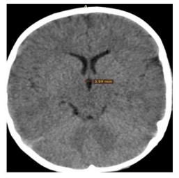 |
| Figure 1: Axial CT image of the brain showing the maximum width of the third ventricle |
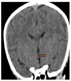 |
| Figure 2:Axial CT image of the brain showing the maximum height of the third ventricle |
Introduction
The human brain is most complex and not yet fully understood organ [1]. The third ventricle is a slit like cleft between the two thalami that may undergo enlargement due to different pathological conditions [3]. It communicates frontally with the lateral ventricles through the interventricular foramina (of Monro) and occipitally with the fourth ventricle through the cerebral aqueduct (of Sylvius) [3].
Ventriculomegaly is a clinically imperative finding that is related with a number of illness. An alter in third ventricle size may be due to its own involvement in a disease process or it can be a manifestation of a disease process elsewhere in the brain parenchyma. Subsequently, it is vital to evaluate ventricular size when doctors assess patients [1]. Evaluation of ventricular size helps in the diagnosis of a disease process or determining the forecast of ventricular shunting [2].
Ventriculomegaly, or expansion of the third ventricle, is a common feature of many neurodegenerative diseases, including Alzheimer’s Disease (AD) and CTE (4). Ventricle expansion in aged subjects with cognitive impairment is strongly correlated to increases in both cognitive decline and altered CSF metabolites that are known biomarkers for AD [4,5]. Ventriculomegaly is also strongly correlated to normal aging [6].
The third ventricle is broadening in an assortment of clinical conditions: such as schizophrenia, hydrocephalus, tumors, and as well as aging which could lead to dementia [6,7]. There was shortage of literary works within the estimation of third ventricle measure in radiology hone of the ranges of the size of cerebral ventricles for the Ethiopian populations. Right now, utilized reference values were drawn from other populaces with races that have diverse epidemiological, statistic and anatomical dissemination.
Radiologists were regularly confronted with issues of choosing whether ventricles are inside ordinary limits or broadened for a patient’s age and sex. This has been a personal decision based on experience [4]. Be that it may, there was bound to be judgmental blunders coming about in misdiagnosis. One such condition that comes about in a require for CT scan evaluation is hydrocephalus.
Our study was pointed to provide computed tomography-based linear measurements of third ventricle of human brain in population of North-west Ethiopia which was lost till date and to discover the regulating information. We attempted here to observe the sexual dimorphism on size of third ventricle in the study population.
Materials and Methods
The cross-sectional and observational computed tomography measurement of third ventricle size was done using cranial CT scans obtained from Department of Radiodiagnosis in University of Gondar comprehensive specialized hospital. The study group comprised of referred patients reporting for CT scan of head region due to various indications. CT scans reported radiologically normal in terms of cerebral ventricles and brain parenchyma were collected in a DVD for analysis.
The study involved 240 ordinary subjects (140 males and 100 females) from 18 to 79 years old, categorized into various groups; 18-30 years, 31-40 years, 41-50 years,51-60 years and above 60 years. CT scan images of head region belonging to study subjects were procured from the Department of Radiodiagnosis. Approval was sought from the Ethics committee of university of Gondar school of medicine some time recently the begun.
All CT scans were performed on 64-slice multidetector spiral CT scanner by trained and experienced radiologists under standardized conditions. CT was done in axial transverse scanning and optimum patient preparation and positioning was taken care of. After obtaining the view in lateral projection (120kVp; 30mA) orbito-meatal line was drawn and a line was drawn at an angle of 15 – 20 degrees to and 1 cm above it, representing the lowest tomographic section, which passed through the base of skull. The sections were taken with slice thickness of 5mm and increment of 10mm. Images were reconstructed at slice thickness of 1.25mm without any overlap.
CT scan images were collected in the DVD with relevant patient information. Analysis of digital images was done on personal laptop, using software tool Radiant DICOM Viewer (64-bit). The measurement was calibrated to nearer 0.1 mm.
Third ventricle was marked as a mid-line structure between right and left thalami. Width of third ventricle was measured in mm (millimeters) at the level of interventricular foramen (foramen of Monro) and was taken as the maximum distance between the two thalami at this level. The height of the third ventricle could not be measured as its posterior marker the pineal gland was not seen in CT scan sections of all patients. The maximum third ventricular width (Figure 1) and the maximum third ventricular height (Figure 2) were measured in mm. The data were entered into Ipinfo, version 7 and analyzed using the IBM SPSS Statistics, version 20. The means (± standard deviation), ranges, minimum, maximum, and the 95% confidence intervals for the mean (in order to include the true population, mean in 95% of the cases) were all calculated. P- Value less than 0.05 is considered as statistically significant.
Results
A total of 240 adults comprising 140 (58.33%) males and 100 (41.67%) females were recruited.
The width of third ventricle reported gradual increment from the age group of 31-40 years to the age greater than 50 years. The maximum mean of third ventricle height was found in the age group of 41-50 years (Table 1).
Two – tailed t-test analysis showed a statistically not significant difference (p>0.05) between third ventricle size of male and female subjects. In both measurements, the mean third ventricle size of male were greater than female (Table 2). The maximum measurement found for the third ventricle height and width in male and female were 18.12cm, 7.32cm and 16.48cm, 6.76 respectively (Table 3).
Pearson`s correlation finding indicated positive correlation between both parameters with age. The TVW and TVH were statistically significant (p 0.05) weak correlation (r˂0.3) with age (Table 4).
Discussion
Present study analyzed computed tomography of 240 patients that were reported radiologically normal; and maximum width and height of third ventricle of brain was evaluated with respect to age and gender in the southwest Ethiopia.
The third ventricle is a slit-like cavity, lies between the right and the left halves of the diencephalon (between two halves of the thalami). It communicates with the lateral ventricles above via interventricular foramina of Monro and with the fourth ventricle below via the cerebral aqueduct of Silvius [2].
Very recent retrospective study conducted on 250 subjects in India reported the mean scores of third ventricle width was (5.8±2.10 mm) [8]. Similarly, the range in the current study of the third ventricle width was between 1.86 to 6.76 millimeters in females, 2.12 to 7.32 millimeters in males and there was a steady rise across age groups until the fifth decade, after which there was a sharp rise. This is because in normal aging ventricles undergo compensatory dilatation with increasing age due to factors such as cortical atrophy, expected feature at this age range [6].
A prospective cohort report of Saudi Arabia revealed that the mean width of third ventricle was 5.57 + 1.60 in 152 subjects (males and females). The width of third ventricle increases as age increases, which was statistically significant (P-value < 0.05) [9].
A prospective study conducted among adult Indian people revealed that the width of the third ventricle was observed to be greater in males (3.47 ± 1.07, 95% CI 3.32 - 3.62 mm) than in females (3.31 ± 0.94, 95% CI 3.16 - 3.46 mm) and this difference was statistically insignificant (T= 1.470 p= 0.164). However, the height of the third ventricle was observed to be greater in females (18.86 ± 8.36, 95% CI 17.52 – 20.21 mm) than in males (17.97 ± 2.76, 95% CI 17.59 -18.35 mm), which was statistically insignificant (T= -1.429 p= 0.154). They found that the third ventricle width and height increase with increase in age of both males and females [10]. On the other hand, report from North West Nigeria indicated that the mean width and height of the third ventricle in the males (5.25 ± 1.32 mm and 27.84 ± 1.53 mm) were greater than those of the females (5.34 ± i1.38 mm and 25.42 ± 1.43 mm) respectively and these differences were statistically significant, P = 0.0209 (< 0.05) [11].
A study conducted in North Indian population revealed that Mean width of third ventricle increased from 2.31 mm to 5.94 mm in males and from 2.51 mm to 5.92 mm in females as the age progressed. Furthermore, maximum width of third ventricle was more in males than females for all the age groups except between 18- 30 years where it was more in females (2.51±1.38 mm) than males (2.31±1.55 mm) though not statistically significant [12].
In our study, we observed third ventricle width and height ranging between 1.86-7.32 mm and 6.85-18.23 mm respectively. Ventricular width was found to be 2.6 mm among subjects who were 31-40 years of age, and it displayed an incremental pattern reaching 3.9 mm in >50 years of age. Width of third ventricle was more in males than females across all age groups. Our findings were in conformation to those of Brinkman et al. (1981) who also reported higher values in males [5].
Conclusion
In this study, it was observed that diameter of third ventricle was 2.98 mm, 2.86 mm in males and females respectively. The third ventricle size were larger in males. The size of third ventricle increased with age. So far, the current study will provide useful information about the normal size of third ventricle while diagnosing hydrocephalus, schizophrenia, psychotic disorders and other pathologies causing ventriculomegaly.
Acknowledgement
We would like to express our deep appreciationto the staff members of Department of Radiology at University of Gondar Comprehensive Specialized Hospital. We wish to acknowledge the medical director/manager of the Universitty of Gondar Comprehensive hospital for his support and permission.
Availability of Data and Materials
The data set supporting this study are available in the manuscript.
Funding
This study received money from the University of Gondar for data collection only. The authors declare that they have received no funds for the publication of this manuscript and that they have no external source of fund for both data collection and publication.
Authors’ Contributions
AA: Conceived the idea, analyze the data, prepare the manuscript: TJ and MJ: supervise the data collection, analyze the data, and prepare the manuscript: all authors approved the manuscript.
Ethical Approval and Consent to Participate
This study obtained ethical approval from College of Medicine and Health Sciences Institutional Review Board of the University of Gondar. The participants gave written informed consent prior to data collection.
Consent for Publication
Not applicable.
Competing Interests
The authors declare that they have no conflict of interests.
References
- Blakemore SJ, Choudhury S (2006) Development of the adolescent brain: implications for executive function and social cognition. Journal of child psychology and psychiatry 47: 296- 312.
- Strominger NL, Demarest RJ, Laemle LB (2012) Gross Anatomy of the Brain. Noback’s Human Nervous System, Seventh Edition: Springer 1-10.
- Marshall LH, Magoun HW (2013) Discoveries in the human brain: Neuroscience prehistory, brain structure, and function: Springer Science & Business Media, USA.
- Kempton MJ, Stahl D, Williams SC, DeLisi LE (2010) Progressive lateral ventricular enlargement in schizophrenia: a meta-analysis of longitudinal MRI studies. Schizophrenia Res 120: 54-62.
- Yadav A, Sharma A, Nigam G, Yadav A, Chauhan K, et al. (2015) Morphometric study of frontal horns of lateral ventricles of the brain by computed tomoraphy in western uttar pradesh population. Journal of Anatomy 23: 22-7.
- Parija B, Sahu N, Rath S, Padhy R (2017) Age-related Changes in Ventricular System of Brain in Normal Individuals Assessed by Computed Tomography Scans. Siriraj Medical Journal 66: 225- 30.
- Styner M, Lieberman JA, McClure RK, Weinberger DR, Jones DW, et al. (2005) Morphometric analysis of lateral ventricles in schizophrenia and healthy controls regarding genetic and disease-specific factors. Proceedings of the National Academy of Sciences 102: 4872-7.
- Honnegowda TM, Nautiyal A, Deepanjan M (2017) A Morphometric Study of Ventricular System of Human Brain by Computerised Tomography in an Indian Population and its Clinical Significance. Austin J Anat 4: 1075.
- Gameraddin M, Alsayed A, Ali A, Al-Raddadi M (2015) Morphometric Analysis of the Brain Ventricles in Normal Subjects Using Computerized Tomography. Open Journal of Radiology 5: 13-9.
- Meshram P, Hattangdi S (2012) The morphometric study of third ventricle and diencephalon by computerized tomography. Indian journal of applied basic medical science 14: 8-13.
- Usman JD, Zagga AD, Tadros AA, Yunusa G, Saidu SA, et al. (2013) Morphological variation of third ventricle using computerized tomography among different gender and age groups: A 5-year retrospective study in Usmanu Danfodiyo University Teaching Hospital, Sokoto, North - West Nigeria. Sahel Med J 16: 83-6.
- Manjari L (2017) Morphometric study of third ventricle width in north Indian population: a CT study. J Anat Science 25: 16-21.
Artcle Information
Review Article
Received Date: May 02, 2025
Accepted Date: May 20, 2025
Published Date: May 27, 2025
Journal of Neuroscience Research and Alzheimer’s Disease
Volume 1 | Issue 1
Citation
Teshome AA, Amare TJ, Taye MJ (2025) Sexual Dimorphism in the Size of Third Ventricle of Human Brain and its Correlation with Age in the Adult Populations of NorthWest Ethiopia, 2019 - An Observational Radiological Study. J Neurosci Res Alzheimers Dis 1: 102
Copyright
©2025 Teshome AA. This is an open-access article distributed under the terms of the Creative Commons Attribution License, which permits unrestricted use, distribution, and reproduction in any medium, provided the original author and source are credited.

doi: jnra.2025.1.102